


Introduction
Type 1 Arnold-Chiari malformation is defined by cerebellar tonsil herniation through the foramen magnum. Although the exact cause of this deformity remains elusive, based on a plausible explanation, the hydrocephalus-driven downward pressure pushes downward various nerve structures located in the posterior fossa, thereby disturbing cerebrospinal fluid outflow.1) The prevalence of congenital type 1 Arnold-Chiari malformation is approximately 1 in 1,000 live births.2) Type 1 Arnold-Chiari malformation symptoms include headache, lower cranial palsy, motor weakness, and ataxia.3) However, isolated dizziness has rarely been described as a first symptom. In this study, we present the case of a type 1 Arnold-Chiari malformation in a previously healthy 14-year-old boy having experienced acute dizziness as a first disease symptom. Moreover, we reviewed previous studies regarding type 1 Arnold-Chiari malformation. Finally, we describe and emphasize the possibility of congenital malformation in the differential diagnosis of dizziness in children.
Case Report
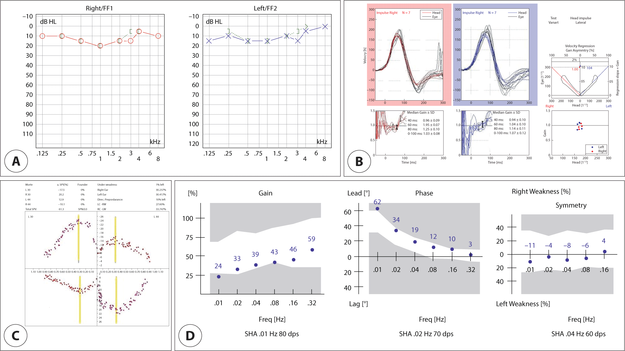
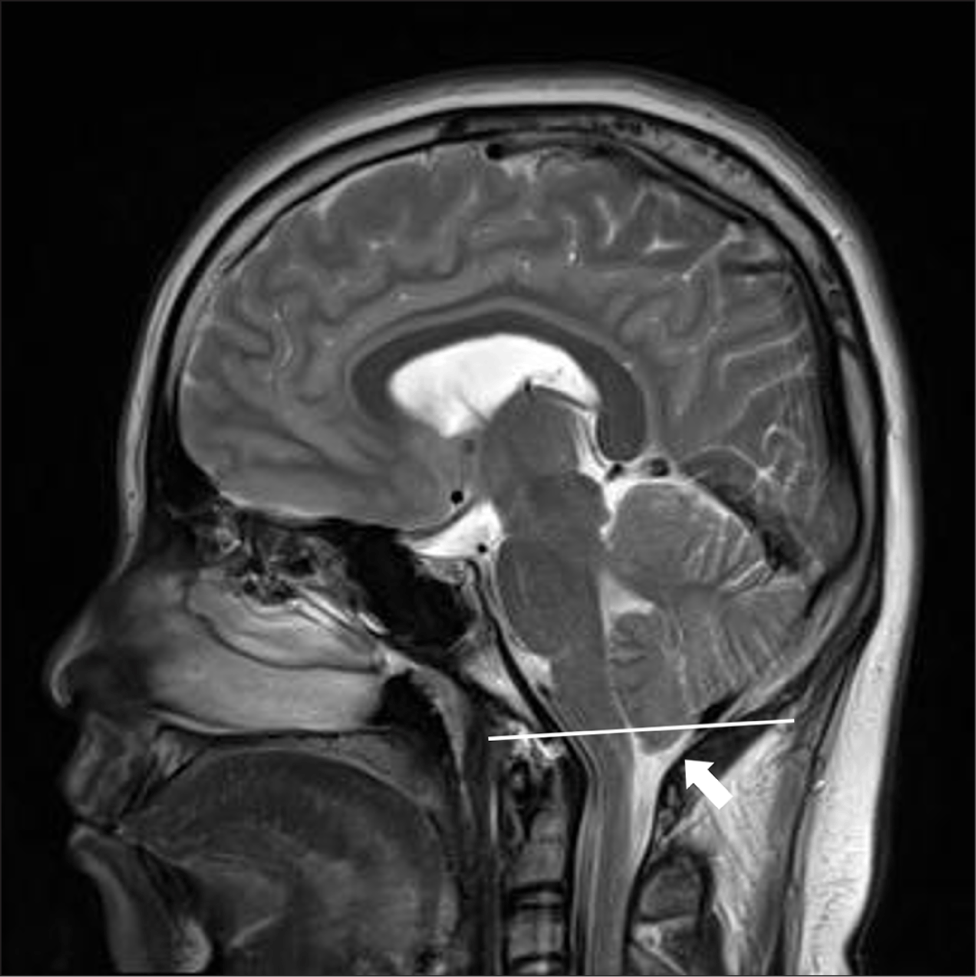
A previously healthy 14-year-old boy was presented to our hospital with a sudden onset of intermittent rotational dizziness, which resolved spontaneously. The clinical presentation is characterized by short attacks of dizziness (several minutes). The patient did not show any signs of vestibular dysfunction between attacks and there were no specific triggering factors. Further, the patient experienced no headache during, before, or after the dizziness. There were no special family and medication history. His symptoms were unrelated to changes in posture and he had no cochlear and autonomic symptoms such as tinnitus or hearing loss and nausea or vomiting, respectively. We observed no spontaneous and gaze-evoked nystagmus on our videonystagmography test. His neurological examination and vestibular functional (i.e., caloric, video head impulse, and rotation chair) test results were normal (Fig. 1). We further consulted with the patient and his parents and conducted further history-taking. Since we registered no underlying anxiety or depressive disorder, we excluded psychiatric dizziness. Vestibular function tests were normal, and if it was not attributed to another disorder, benign paroxysmal vertigo of childhood had to be considered. To assess the potential central origin of dizziness, we performed brain magnetic resonance imaging (MRI). On the sagittal T1 image, we observed that the cerebellar tonsils were located more than 5 mm below the occipital foramen, thereby we confirmed herniation (Fig. 2) and, based on these radiological findings, we diagnosed the patient with type 1 Arnold-Chiari malformation. Following diagnosis, the patient received a cervical collar to limit cervical movement and prevent the worsening of the symptoms. Since dizziness was not severe, the patient was closely followed up in an outpatient clinic without surgical treatment. After a 1-year follow-up, the patient presented no worsening of the dizziness symptoms.
Discussion
Although Arnold-Chiari malformation is a disease with very diverse etiologies and clinical manifestations, it displays one characteristic in common: the malformation of cerebellar and brainstem anatomical structures. Arnold-Chiari malformation is classified as type I–IV depending on the anatomical malformation degree of the cerebellum, brainstem, and craniocervical junction, types III and IV being very rare and accompanied by serious malformations.4) Concerning type I, although symptoms might appear during childhood, in several cases, they appear in the 30s or 50s without any symptoms.3) Type 1 Arnold-Chiari malformation could be diagnosed by radiological examination, when the MRI indicates a cerebellar tonsil protrusion beyond 5 mm from the occipital foramen, along with bony deformities in the posterior fossa and the craniocervical junction. Symptoms such as pain as well as reduced motor skills and sensation might occur, and headache is reportedly the most common symptom. In addition, central cord syndrome, leading to movement and sensation loss in the upper extremities, might occur. Foramen magnum compression syndrome could also be present with potential abnormalities in the corticospinal tract, reduced sensation, cerebellar dysfunction, and lower cranial nerve palsies, with 80% of the cases developing ocular symptoms (e.g., nystagmus, double vision, and otalgia).5)
The treatment of Arnold-Chiari malformation is mainly divided into observation and surgical decompression. In the case of mild symptoms, follow-up observation could be considered with neck movement restriction using a cervical collar to prevent the worsening of the symptoms.6) Decompression surgery is an option in cases of nerve paralysis, myelopathy, cerebellar symptoms, severe neck pain, and occipital headache.
In the presented case, the patient complained of intermittent rotational dizziness with no headache. In addition, his physical signs were not accompanied by motor defects or reduced sensation. Since we observed no specific findings in the vestibular function and hearing tests, we performed a brain MRI to differentiate dizziness of central origin such as cerebellar lesions, and diagnosed the patient with type 1 Arnold-Chiari malformation.
Dizziness in children is of common occurrence in recent clinical practice with a reported prevalence of approximately 15%.7) The most common dizziness-related diseases in children comprise vestibular migraine, benign paroxysmal vertigo in childhood, somatoform disorder, and vestibular neuritis. Dizziness of central origin is due to lesions in the brainstem (vestibular nuclei, ocular motor nuclei, medial longitudinal fascicle, reticular formation, midbrain tegmentum) and in the cerebellum (flocculus and nodulus). Supratentorial lesions (thalamus, cortex) rarely cause rotatory vertigo, but affected patients can present with dizziness and deviation of the subjective vertical.8) Tumors of the brainstem and the cerebellum can also cause vertigo in children. MRI should be performed if the clinical examination reveals central ocular motor signs.8) The characteristics of the dizziness-causing disease itself in children do not differ significantly from those in adults.9,10) However, congenital malformations and epileptic vertigo might occur at a higher frequency in children than in adults, evaluating these conditions is thus essential.11) If the symptoms or non-specific dizziness display no improvement, similar to the hereby presented case, dizziness of central origin must be identified.