


Introduction
The injection of anesthetics, corticosteroids, and botulinum toxin into muscles, as well as dry needling, commonly referred to as trigger point injection (TPI), is a widely employed approach for alleviating myofascial pain. TPI typically targets the most sensitive and irritable muscle points identified through palpation, with or without ultrasound (US) guidance.1–3) The posterior neck muscles are common sites for TPI to alleviate cervical myofascial pain or chronic headaches.1) Due to the concurrent involvement of various posterior neck muscles in the development of cervical pain and headache, TPIs are often administered simultaneously to multiple muscles.4)
Skin or soft tissue infections are among the most common complications associated with TPI.5) While infections due to TPI typically resolve with conservative treatments such as antibiotic therapy, some cases necessitate surgical drainage of abscesses to control infectious conditions effectively. In this report, we describe our experience with draining abscesses that developed in the splenius capitis (SC) and levator scapulae (LS) muscles after TPI, which are anatomical locations less familiar to head and neck surgeons. Additionally, we provide a review of key considerations during the surgical drainage process.
Case Report
An 18-year-old healthy man presented to the emergency room with worsening left posterior neck pain, persisting for eight days. He had undergone multiple injections to alleviate pain in the left posterior neck muscle four days prior to the visit. The pain intensified rapidly on the next day of TPI, accompanied by fever and swelling in the left neck. Physical examination revealed severe tenderness of the left posterior neck and swelling of the left posterior pharyngeal wall (Fig. 1A, B). However, there were no neurological symptoms suggestive of peripheral neuropathy, such as limited arm motion or radiating pain. Laboratory tests showed neutrophil-dominant leukocytosis (14.12×10³ cells/μL, [reference: 4.0×10³–10.0×10³ cells/μL]) and elevated C-reactive protein levels (8.44 mg/dL, [reference: 0–0.5 mg/dL]).

Contrast-enhanced computed tomography (CT) revealed a 15 mm×14 mm×13 mm sized abscess with central low attenuation and surrounding rim enhancement in the left SC muscle between the C2 and C4 vertebrae. The abscess extended to the left side of the retropharyngeal space (Fig. 2A–C), with the V2 segment of the vertebral artery (VA) located close to its medial side (Fig. 2A, B). Furthermore, fluid collection around the abscess extended medially to the paraspinal muscles and inferiorly to the LS muscles.
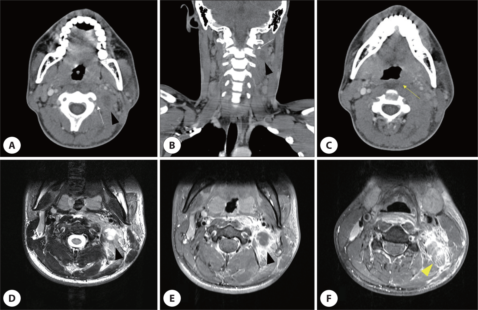
Magnetic resonance imaging (MRI) was performed to evaluate the relationship between the spinal lesion and the abscess. MRI revealed an abscess in the left SC muscle at the level of C2–C4 vertebrae, exhibiting low signal intensity on T1-weighted imaging and high signal intensity on T2-weighted imaging (Fig. 2D, E). In addition, a high signal around the lower portion of the LS muscle was only observed on T2-weighted imaging indicating fluid collection (Fig. 2F). No spinal cord lesions were observed.
After blood cultures were conducted to identify the pathogen, intravenous vancomycin (15 mg/kg every 12 hours) was initiated as empirical antibiotic therapy for abscesses against potential methicillin-resistant Staphylococcus aureus, as recommended by the Infectious Diseases Department.
Due to the rapid worsening of neck swelling and pain, the SC muscle abscess was drained under general anesthesia. Using US in the prone position, the location of the abscess and major vascular systems, especially the VA located medial to the abscess, was delineated. Following skin incision, the anterior border of the upper left trapezius muscle was identified, and dissection was continued to confirm the SC muscle. The abscess was drained without any major bleeding events, and a Jackson-Pratt drain was inserted under negative pressure. Additionally, drainage of the left retropharyngeal abscess was performed using a transoral approach.
Following surgery, the patient’s upper posterior neck pain and swelling significantly improved, with no specific pathogens detected in blood or pus cultures. Vancomycin administration was continued after consultation with the Infectious Diseases Department.
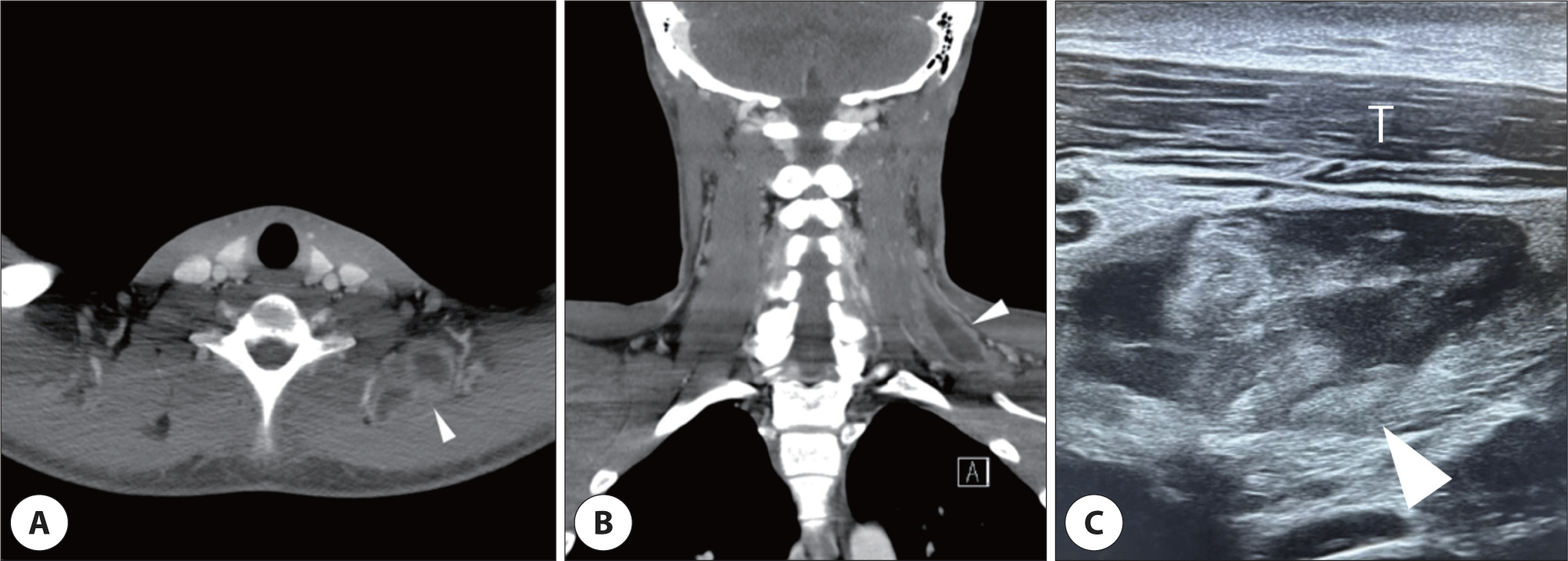
During the physical examination on postoperative day five, the patient complained of left shoulder pain in the lower portion of the upper trapezius muscle, which gradually worsened until postoperative day nine. A CT scan revealed a newly formed abscess within the LS muscle between the level of C6 and T1 vertebrae, where signal changes were observe at the initial presentation (Fig. 3A, B). US revealed a well-defined abscess in the LS muscle beneath the trapezius (Fig. 3C). Despite daily repeated aspirations guided by US, there was no significant improvement, prompting drainage of the abscess through the lower portion of the posterior triangle of the neck.
Intraoperative nerve monitoring of the spinal accessory nerve (SAN) was performed before incision to prevent injury, considering the possibility of encountering its entry point into the trapezius muscle, which is generally situated 2–4 cm above the clavicle. While approaching the LS muscle, the SAN was identified and successfully preserved without injury. With symptom improvement following abscess drainage, the patient was discharged on postoperative day six, and no signs of recurrence were observed during subsequent outpatient follow-ups.
Discussion
Although TPI of the neck muscles is not commonly practiced by otolaryngologists, it has been widely used in pain medicine by other medical professionals to target various skeletal muscles of the neck and face, including the masseter, SC, LS, trapezius, and sternocleidomastoid (SCM) muscles.1–3) TPI delivers drugs into the muscle or fascia through an aseptic procedure. However, inflammation induced by TPI can occur due to infection or muscle necrosis following steroid injection. Infection should be considered in patients with increased neck pain after TPI, as observed in this case. Invasive interventions by head and neck surgeons may occasionally be necessary to treat TPI complications. We successfully drained abscesses caused by TPI in the SC and LS muscles, which are rarely encountered in otorhinolaryngology.
Antibiotics are usually the primary treatment choice for TPI-induced infections. However, despite antibiotic treatment, surgical drainage should be promptly considered in patients with progressive infection or presenting significant symptoms such as extreme pain, high fever, or complex abscess formation, to prevent septicemia.6–9) In cases without significant symptoms, less invasive treatments can be considered. Since previous studies have reported comparable efficacy of US-guided repeated aspiration to surgical drainage for intramuscular abscesses, US-guided repeated aspiration can be a reasonable treatment option.6,8) However, caution is warranted when the abscess is near the major vasculature or critical structures during US-guided aspiration, especially when performed by an inexperienced clinician.6)
The SC muscle, known to head and neck surgeons as the landmark for level IIb dissection, originates from the spinous processes of the T3–T5 vertebrae and inserts at the transverse processes of the C1 and C2 vertebrae, superior nuchal line, mastoid process, and occipital bone.10) It lies deeper than the trapezius and LS muscles, and is a common target for TPI because it is known to cause cervical pain and chronic headache.1) When approaching the upper portion of the SC muscle, the course of the VA should be clearly identified. The VA, especially the V2 segment, ascends through the transverse foramen from the C6 to the C2 vertebrae.11) Subsequently, it exits from the C2 vertebra to the posterior neck muscle, which is located deeper than the SC muscle within the suboccipital triangle.12,13) The course of this artery may vary between individuals until it reaches the C1 vertebra.12) To prevent VA damage and specify SC muscle location, it is necessary to expose the posterior margin of the SCM muscle and identify the posterior neck triangle.12,14)
Furthermore, it is important to be cautious of potential damage to the occipital artery (OA), a major vasculature when exposing the SC muscle. The OA, a branch of the external carotid artery, is initially covered by the SCM and SC muscles and travels backward along a groove in the temporal bone near the mastoid process.15) Before exposing the SC muscle through the anterior border of the trapezius muscle, utilizing US to delineate the OA can minimize the risk of injury. The course of the SAN in the upper posterior triangle of the neck is also critical when exposing the SC muscle. Limited anterior dissection from the anterior border of the trapezius muscle should be performed, considering the specific distance between the anterior border of the trapezius muscle and SAN in the upper posterior triangle of the neck.16)
The LS muscle, extending from the C1 to T2 vertebrae and located deep beneath the trapezius muscle, is another common target for TPI.17) In this case, the initial MRI revealed a high T2 signal change without a low T1 signal in the lower portion of the LS muscle, likely resulting from either the initial injection or needle penetration into the LS muscle during multiple TPIs on the trapezius or SC muscles.10) Since there was no definitive abscess formation in the LS muscle at the initial presentation, antibiotic treatment could be considered. However, due to the progressive nature of the abscess and the need for invasive treatment, we conducted repeated US-guided aspirations.6)
Despite repeated aspirations, the LS muscle abscess continued to progress, necessitating surgical drainage. When approaching the lower portion of the LS muscle, where the abscess was located in this patient, surgeons should be mindful of potential injury to the SAN.17) During abscess drainage in the LS muscle by exposing it, surgeons may encounter the SAN.18,19) Therefore, consideration should be given to nerve monitoring to prevent SAN injury and ensure its identification when draining abscesses in the lower portion of the LS muscle.19)
Furthermore, when approaching the LS muscle, surgeons should be cautious regarding potential injury to the nerve to the LS muscle (LSN), originating from the C4 root of the brachial plexus.19) Considering the course of this nerve, attention should be paid to potential injuries if a clear inspection reveals the LS-overlying fascia. Nerve monitoring of the brachial plexus or a cautious approach to the LS muscle along the anterior border of the trapezius muscle can help prevent LSN injury.
Conclusion
Head and neck surgeons sometimes require abscesses in the posterior neck muscle following TPI. Given the unfamiliarity of posterior neck anatomy to head and neck surgeons, preventing damage to major anatomical structures, especially the VA and OA in the upper portion of the SC muscle, as well as the SAN and LSN in the lower portion of the LS muscle, is crucial. Ancillary techniques, such as nerve monitoring and US-guided identification, may be helpful in preventing damage to these structures.