증례

경비강 내시경 수술을 이용한 재발성 비인두암의 치험 1예

서윤수1, 조기헌1, 이형신1,*
Yoon Soo Seo1, Ki Hun Jo1, Hyoung Shin Lee1,*
Author Information & Copyright
1고신대학교 의과대학 이비인후-두경부외과학교실
1Department of Otolaryngology-Head and Neck Surgery, Kosin University College of Medicine, Busan, Korea
*교신저자: 이형신, 49267 부산광역시 서구 감천로 262 고신대학교 의과대학 이비인후-두경부외과학교실 전화 :(051) 990-6470·전송 :(051) 245-8539 E-mail:sego78@hanmail.net

© Copyright 2019 The Busan, Ulsan, Gyeoungnam Branch of Korean Society of Otolaryngology-Head and Neck Surgery. This is an Open-Access article distributed under the terms of the Creative Commons Attribution Non-Commercial License (http://creativecommons.org/licenses/by-nc/4.0/) which permits unrestricted non-commercial use, distribution, and reproduction in any medium, provided the original work is properly cited.

Received: Aug 12, 2019; Revised: Oct 11, 2019; Accepted: Nov 19, 2019

Published Online: Dec 31, 2019

ABSTRACT

A 59-year-old male treated with radiotherapy presented with recurrent nasopharyngeal mass during the follow-up period of 21 months. Recurrence was confirmed by biopsy. The recurrence was relatively localized and no regional or distal metastasis was observed by imaging study. Transnasal endoscopic excision of tumor was performed with safety margin of 0.5 cm. The patient was discharged 1 day after surgery without any complication and there was no recurrence during the follow-up period of 2 years. In this case, we propose that surgical removal by transnasal endoscopic approach is feasible for treatment of early-stage recurrent nasopharyngeal cancer. (J Clinical Otolaryngol 2019;30:294-297)

Keywords: 재발성 비인두암; 내시경적 절제술
Keywords: Recurrent nasopharyngeal carcinoma; Endoscopic resection

서 론

비인두암은 비인두부의 상피에 발생하는 악성종양으 로 증상이 없거나 비출혈과 같은 비특이적 증상을 보여 늦게 발견하는 경우가 많아 예후가 좋지 않은 경향을 보 인다.1,2) 10만명당 15~30명의 발병률을 보이는 암으로 방 사선 치료에 비교적 잘 반응하기 때문에 주 치료로서 방 사선 단독요법 또는 항암방사선병합요법을 고려하게 된 다.3-6) 초기인 경우 방사선 치료 단독으로 완치가 가능한 데 이 경우 방사선 치료 후 5년 생존률은 85~90% 정도로 좋은 예후를 보이나 8~10% 정도에서는 재발을 경험하며 이들 중에는 원격전이가 발생하는 경우도 흔하다.5,6) 조 기 비인두암의 좋은 치료성적에 비하여 재발한 비인두 암의 경우 발견이 어렵고 진행된 병변으로 확인되는 경 우가 많아 좋지 않은 예후를 보인다.1,5,7) 재발한 비인두 암의 치료로는 방사선 치료의 재시도나 항암요법을 시 도해 볼 수 있으며 비인두부위에 병변이 국한되어 있는 조기 재발성 비인두암의 경우에는 수술적 절제를 제한 적으로 고려할 수 있다.4-6) 본 증례의 경우 정기적인 내시 경 검사를 통해 조기에 발견한 재발성 비인두암을 침습 적인 절개 또는 방사선치료의 재시도 없이 경비강 내시 경으로 제거하여 좋은 결과를 얻어 보고 하고자 한다.

증 례

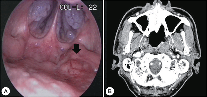
59세 남자가 간헐적 비출혈을 주소로 본원 외래로 내 원하였다. 과거력 상 고혈압, 신증후군이 있었다. 음주력 은 주 1회 맥주1병 정도였고, 15갑년의 흡연력 있었다. 비인두내시경에서 좌측 비인두부에 경계가 불분명한 병 변이 관찰되었고(Fig. 1A), 종괴는 비인두강내 국한되어 있었다. 경부 이학적 검사상 구인두 및 비강에 특이소견 은 없었고, 경부 촉진에서도 종물은 만져지지 않았다. 경 부 전산화단층촬영에서 좌측 귀인두관 융기 내측에 0.6 × 0.6 cm 크기의 조영증강되는 종물이 관찰되었으며 (Fig. 1B), 경부 림프절 전이는 관찰되지 않았다. 펀치 생 검을 통한 조직검사 결과는 비각화 편평상피암종으로 비인두 암(T1N0M0, stage I)으로 진단하였다. 초 치료로 방사선 치료가 6840 cGy(38회) 시행되었다.
jcohns-30-2-294-g1
Fig. 1. Image of nasopharyngeal cancer before radiotherapy. A : Nasopharyngoscopy image before radiotherapy shows a protruded mass at left nasopharynx (arrowed). B : Contrast-enhanced computerized tomography axial view before radiotherapy shows an enhanced mass at left nasopharynx.
Download Original Figure

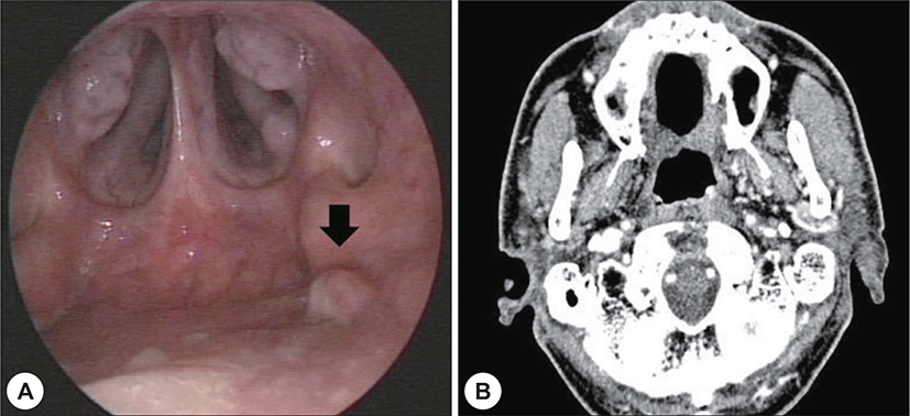
치료 21개월 후 비인두강 내시경상 좌측 비인두부에 경계가 불분명한 병변이 관찰되었고(Fig. 2A) 종양의 조 직검사 결과 저분화성 비각화 편평상피암종으로 확인되 어 재발성 비인두암으로 진단하였다. EBV면역조직검사 에서는 음성으로 확인되었다. 병기확인을 위한 PET 전 산화 단층촬영에서는 저명한 병변이 확인되지 않았고 경부 림프절이나 타 장기로의 전이 소견 또한 관찰되지 않았다.
jcohns-30-2-294-g2
Fig. 2. Image of nasopharyngeal cancer after recurrence. A : Nasopharyngoscopy 2 years after radiotherapy shows a recurrent mass (arrowed). B : Contrast-enhanced computerized tomography axial view after recurrence shows an enhanced mass at left nasopharynx without significant submucosal invasion.
Download Original Figure

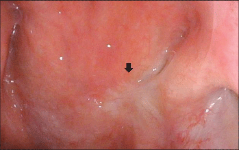
경부 전산화 단층촬영 영상에서 병변이 비교적 국한되 어 있고 점막 하로 깊이 침윤하는 소견이 관찰되지 않아, 경비강 내시경 수술을 시행하기로 하였다(Fig. 2B). 충분 한 수술 공간을 확보하기 위하여 양측 하비갑개 외향골 절술을 시행 하였으며 수술 기구의 충분한 가동범위를 확보하기 위하여 보조의가 우측 비강으로 내시경을 삽 입하여 좌측 비인두의 병변부위를 확인하였으며(Fig. 3A) 시야를 충분히 확보한 상태에서 좌측 비강으로 흡 입기나 내시경 겸자를 삽입하여 병변부위의 조직을 견 인하면서 우측 비강으로 삽입한 단극성 소작기를 이용 하여 병변의 절제범위를 표시하였다(Fig. 3B). 안전연은 0.5 cm으로 설정하였으며 단극성 소작기를 이용하여 상 인두수축근까지 안전변연을 확보하여 충분한 깊이로 병 변부위를 제거하였고(Fig. 3C) 1.6×1.6×2.0 cm 크기의 조직을 절제하였다. 이후 출혈부위를 양극성 소작기를 이용하여 지혈 후 수술을 마쳤으며 환자는 수술 1일째 특 별한 합병증 없이 퇴원하였다. 조직검사결과 0.6×0.8 cm 크기의 편평상피세포암으로 확인되었으며 절제변연은 음성으로 확인되었고 환자는 수술 2년 후 비인두내시경 및 영상학적 검사에서 암의 재발 없이 경과관찰 중이다 (Fig. 4).

jcohns-30-2-294-g3
Fig. 3. Surgical endoscopic image. A : Visibility was obtained by inserting an endoscopy into opposite nasal cavity. B : Excision was conducted with a unipolar cauterization while pulling the tissue using a suction tip. C : Completion of operation after hemostasis with electrocautery.
Download Original Figure
jcohns-30-2-294-g4
Fig. 4. Nasopharyngoscopy image of postoperative 2 year. Image shows no evidence of disease with post-operative scar (arrowed).
Download Original Figure

고 찰

비인두암의 치료 방법은 병기에 따라 결정하는데, 방 사선 치료의 효과가 좋은 암종이므로 조기암의 경우 방 사선치료를 주로 시행하며 진행암의 경우 항암방사선병 합요법을 시행한다.5) 원발암의 수술적 절제는 재발암인 경우에 제한적으로 고려하게 된다.5,8) T1~2의 조기 비인 두암은 방사선 단독치료에 대한 국소반응률이 양호한 질 환이나 8~10.9% 정도에서 재발하게 되는데 재발한 비인 두암은 방사선치료에 저항성을 가질 수 있어 치료방법 의 선택이 제한적이며 50% 이상에서 진행된 병기로 발 견되기 때문에 좋지 않은 예후를 보일 수 있어 재발을 조 기에 발견하는 것이 중요하다.3,5-7,9)

일반적으로 방사선 치료에 대한 반응이 없는 경우 수 술적 절제를 고려해 볼 수 있으며, 전통적인 수술방법으 로 경상악 접근법과 경구개접근법, 경하악-익상접근법 이 있다.4,5,10) 이러한 수술은 비인두부로 접근을 위한 침습 적인 수술로 익상근의 손상 등의 이차적 손상으로 인한 개 구장애 및 삼킴장애와 안면변형 등의 가능성이 있다.4,10)

침습적인 수술적 치료에 비하여 경비강 내시경적 비인 두절제술은 조기 병변이나 국소 재발한 비인두암에서 시 행할 수 있는 최소 침습적인 수술적 접근법으로 좁은 비 강으로 인하여 구획 내 절제가 쉽지 않으며 비인두부의 괴사나 출혈, 과대비성과 같은 합병증이 발생할 수 있다 는 단점이 있으나 경상악 접근법 등의 침습적 접근법 보 다 시야확보에 용이한 이점이 있다.3) 또한 수술적 합병증 이 비교적 적으며 조직의 손상을 줄일 수 있고 회복기간 이 더 빠르다는 장점이 있다.5,11-13) 그리고 전통적인 접근 법에 비하여 경동맥과 같은 주요 구조물의 노출을 최소 화 할 수 있어 심각한 합병증의 가능성을 줄일 수 있다.10) 경비강 내시경수술을 이용한 재발성 비인두암 치료 후 2년 생존률은 80~100%, 국소 재발률은 12%로 보고되고 있다.5,10-14)

하지만 국내에서 재발성 비인두암의 경비강 내시경적 절제에 대한 보고는 없다.

본 증례에서는 방사선 치료 후 정기적인 비인두내시 경 검사를 시행하여 재발성 비인두암의 조기발견이 가 능하였다.13) 경비강 내시경을 이용한 시야 확보 및 종양 의 구획 내 절제를 시행하였으며 특별한 수술 합병증 없 이 퇴원하였고 이후 2년의 추적관찰 동안 재발소견은 보 이지 않았다. 비인두암의 초 치료 후 내시경을 이용한 정 기적인 추적관찰로 재발암이 초기에 발견되면 경비강 내시경 수술을 통하여 비교적 간단하고 안전하게 병변 을 절제할 수 있으므로 이를 재발성 비인두암의 치료방 법으로 고려해 볼 수 있겠다.

REFERENCES

1).

Lee HM, Okuda KS, Gonzalez FE, Patel V. Current perspectives on nasopharyngeal carcinoma. Adv Exp Med Biol 2019;1164:11-34.

2).

Paul P, Deka H, Malakar AK, Halder B, Chakraborty S. Nasopharyngeal carcinoma: understanding its molecular biology at a fine scale. Eur J Cancer Prev 2018;27(1):33-41.

3).

Xu C, Chen YP, Ma J. Clinical trials in nasopharyngeal carcinoma-past, present and future. Chin Clin Oncol 2016; 5(2):20.

4).

Yoshizaki T, Ito M, Murono S, Wakisaka N, Kondo S, Endo K. Current understanding and management of nasopharyngeal carcinoma. Auris Nasus Larynx 2012;39(2):137-44.

5).

Perri F, Della Vittoria Scarpati G, Caponigro F, Ionna F, Longo F, Buonopane S, et al. Management of recurrent nasopharyngeal carcinoma: current perspectives. Onco Targets Ther 2019;12:1583-91.

6).

Chua MLK, Wee JTS, Hui EP, Chan ATC. Nasopharyngeal carcinoma. Lancet 2016;387:1012-24.

7).

Teo PT, Tan NC, Khoo JB. Imaging appearances for recurrent nasopharyngeal carcinoma and post-salvage nasopharyngectomy. Clin Radiol 2013;68(11):e629-38.

8).

Kontny U, Franzen S, Behrends U, Bührlen M, Christiansen H, Delecluse H, et al. Diagnosis and treatment of nasopharyngeal carcinoma in children and adolescents - recommendations of the GPOH-NPC study group. Klin Padiatr 2016;228(3):105-12.

9).

You R, Zou X, Hua YJ, Han F, Li L, Zhao C, et al. Salvage endoscopic nasopharyngectomy is superior to intensity-modulated radiation therapy for local recurrence of selected T1-T3 nasopharyngeal carcinoma-A case-matched comparison. Radiother Oncol 2015;115(3):399-406.

10).

Chen MY, Wen WP, Guo X, Yang AK, Qian CN, Hua YJ, et al. Endoscopic nasopharyngectomy for locally recurrent nasopharyngeal carcinoma. Laryngoscope 2009;119(3):516-22.

11).

Stoker SD, van Diessen JN, de Boer JP, Karakullukcu B, Leemans CR, Tan IB. Current treatment options for local residual nasopharyngeal carcinoma. Curr Treat Options Oncol 2013;14(4):475-91.

12).

Huang Y, Qiu QH. Endoscopic surgery for early-stage nasopharyngeal carcinoma: a justified initial option. Acta Otolaryngol 2017;137(11):1194-8.

13).

Liu J, Yu H, Sun X, Wang D, Gu Y, Liu Q, et al. Salvage endoscopic nasopharyngectomy for local recurrent or residual nasopharyngeal carcinoma: a 10-year experience. Int J Clin Oncol2017;22(5):834-42.

14).

Zou X, Han F, Ma WJ, Deng MQ, Jiang R, Guo L, et al. Salvage endoscopic nasopharyngectomy and intensity-modulated radiotherapy versus conventional radiotherapy in treating locally recurrent nasopharyngeal carcinoma. Head Neck 2015;37(8):1108-15.