Journal of Clinical Otolaryngology Head and Neck Surgery
The Busan, Ulsan, Gyeoungnam Branch of Korean Society of Otolaryngology-Head and Neck Surgery
Original Article

한국인의 내시경 부비동 수술에서 Keros와 Gera 분류 간의 상관관계 검증

손상준1https://orcid.org/0000-0001-6403-5278, 구형본1https://orcid.org/0000-0002-1342-9450, 윤기서1https://orcid.org/0009-0002-4991-4041, 이재훈1,*https://orcid.org/0000-0002-0856-8173
Sang-Jun Son1https://orcid.org/0000-0001-6403-5278, Hyung-Bon Koo1https://orcid.org/0000-0002-1342-9450, Kiseo Yoon1https://orcid.org/0009-0002-4991-4041, Jae-Hoon Lee1,*https://orcid.org/0000-0002-0856-8173
1원광대학교 의과대학 이비인후과학교실
1Department of Otolaryngology-Head and Neck Surgery, Wonkwang University School of Medicine, Iksan, Korea
*Corresponding author: Jae-Hoon Lee, Department of Otolaryngology-Head and Neck Surgery, Wonkwang University School of Medicine, Iksan 54538, Korea, Tel: +82-63-859-1441, Fax: +82-63-841-6556, E-mail: Leejaehoon64@gmail.com

© Copyright 2024 xxxxxxxxxxxxxxx. This is an Open-Access article distributed under the terms of the Creative Commons Attribution Non-Commercial License (http://creativecommons.org/licenses/by-nc/4.0/) which permits unrestricted non-commercial use, distribution, and reproduction in any medium, provided the original work is properly cited.

Received: Jun 27, 2024; Revised: Aug 13, 2024; Accepted: Sep 26, 2024

Published Online: Dec 31, 2024

ABSTRACT

Background and Objectives:

The lateral lamella of cribriform plate is the thinnest part of the skull base (SB), and there is a possibility of perforation during endoscopic sinus surgery (ESS). Keros and Gera classifications are known for predicting the risk of damage to the SB. The Keros classification is divided into three types according to the depth of the olfactory fossa, and the longer the depth of the olfactory fossa, the more vulnerable it is to damage SB. The Gera classification was divided into three types by measuring the slope of the ethmoid roof as an angle, and the smaller the angle, the more vulnerable it is to damage the SB. Therefore, this study attempted to analyze the correlation between the both classifications in Koreans.

Materials and Methods:

We analyzed 294 adult patients with sinus computed tomography images (588 sides) performed for diagnosis. We measured the lateral lamella length (LLL, distance between the fovea ethmoidalis and the horizontal cribriform plate in the olfactory fossa) and Gera angle (GA, angle between LLCP and lateral extension of horizontal plane passing through cribriform plate), and classified them into each type. Statistical analysis was performed for using the chi-square test, student t test, paired t test, and Pearson's correlation test.

Results:

There was no gender difference in the LLL (p=0.406). The GA in females (64.0±12.3°) was greater than that in males (61.3±14.0°), showing significant gender difference (p=0.019). The average LLL on the left side (5.92±1.92 mm) was significantly longer than that on the right (5.49±1.79 mm) (p<0.001). The average GA on the left side (63.3±13.1°) was significantly larger than that on the right (61.8±13.5°; p=0.017). The LLL and GA showed a strong positive correlation (r=0.629, p=<0.001).

Conclusion:

The significant differences of LLL and GA between both sides and a significantly higher GA in female than male patients raises a precaution for rhinology surgeons to more appreciate the inter-personal and gender-differences of the anterior cranial fossa SB anatomy upon performing ESS or SB surgery.

Keywords: Endoscopic sinus surgery; Skull base; Lateral lamealla; Cribriform plate; Keros; Gera; Classification

서론

사판측벽(lateral lamella of cribriform plate)은 뇌 기저부에서 가장 얇은 부위로 내시경 부비동 수술 과정에서 천공 발생 가능성이 있다.1,2) 사판측벽 길이는 후각와(olfactory fossa)의 깊이에 의해 평가된다. 1962년에 Keros는 후각와의 깊이를 I형은 1–3 mm(저위험), II형은 4–7 mm(중위험) 그리고 III형은 7 mm 이상(고위험)으로 분류하였다.3) 후각와의 깊이가 깊을수록 사판측벽의 의인성 손상 위험성이 증가하며 III형이 가장 취약한 유형이다. 현재까지도 Keros 분류는 사판측벽 길이(후각와 깊이)에 따라 뇌 기저부 손상의 위험성에 대한 평가 방법으로 널리 사용되어 왔다.

최근에는 수평사판(horizontal cribriform plate)을 통과하는 수평면의 측벽연장선과 사판측벽 사이의 각도에 따른 Gera 분류가 제안되었다.4) 뇌 기저부 손상위험도는 80°보다 초과되는 타입 I은 저위험군이고, 45° 이상 80° 이하인 타입 II은 중위험군, 45° 미만인 타입 III은 고위험군에 해당된다. 각도가 작을수록 즉, 타입 III가 손상위험도가 가장 크다.

뇌 기저부에 대한 손상 위험을 예측하는 데 대표적인 두 분류인 Keros 분류와 Gera 분류의 상관성에 대해 국내의 보고는 없었으며, 소수의 국외 연구자들에 의해 연구들이 진행되어 왔으나 서로 상이한 결과를 보였다.4-6) 그래서 본 연구는 한국인을 대상으로 두 분류를 시행하고, 두 분류 간의 상관관계에 대한 분석을 한국인에서 처음 시행하고자 하였다. 그리고 추가적으로 두 분류를 통해 연구군의 성별 간 혹은 좌, 우측 사이에 차이를 보이는지 분석하고 기존 연구들과 비교 및 고찰하여 본 연구의 분석이 내시경 부비동 수술에 임상적인 중요성이 있을지 확인하고자 하였다.

대상 및 방법

대상

2017년 7월부터 2018년 12월까지 본 병원에 비부비동염 증상으로 내원하여 부비동 단층촬영(computed tomograph, CT)을 시행한 후 비부비동염 유무를 확인했던 18세에서 70세까지 총 294명을 대상으로 후향적 연구를 수행하였다. 연구대상군의 선정 기준은 부비동 CT에서 비부비동 병변이 없거나, 비부비동 병변이 있어 약물치료 혹은 수술적 치료를 받았으나 본 연구의 영상학적 계측에 영향이 없는 경우로 하였다. 제외 기준으로는 비부비동 병변이 영상학적 계측에 혼돈을 줄 수 있는 다발성 용종, 종양성 및 낭종성 병변, 그리고 코 수술 기왕력 혹은 과거 안면 골절이 있는 경우였다. 본 연구는 본 병원 기관심의위원회의 승인(2023-05-009)을 받고 수행되었다.

고해상력 부비동 단층촬영을 통한 분석

294명의 연구대상군에서의 부비동 단층촬영은 Somatom Definition Flash 256-slice CT scanners(Simens Healthineers, Forchheim, Germany)를 이용하여 1 mm 두께로 축상면을 측정하였으며, 관상면과 시상면은 Wizard workstation(Simens Healthineers)을 통해 2 mm 두께로 재구성되었다. 좌, 우측을 포함한 총 588측을 분석하였다.

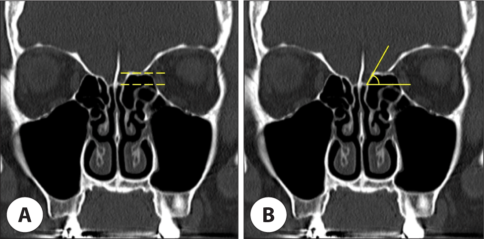
Keros 분류에서 사판측벽 길이의 측정은 Koo 등7)의 방법을 참고하여 관상면들 가운데 볏돌기(crista galli)가 잘 관찰된 부분을 선택하여 후각와에서 사골와(fovea ethmoidalis)의 수평선과 수평 사판(horizontal cribriform plate)의 수평선 사이의 수직 거리를 측정하였다(Fig. 1A). 사판측벽 길이에 따라 I형은 1–3 mm(저위험), II형은 4–7 mm(중위험) 그리고 III형은 7 mm 이상(고위험)으로 구분하였다.
jcohns-35-4-145-g1
Fig. 1. Lateral lamella length and Gera angle. The lateral lamella length is measured as the distance between the fovea ethmoidalis and the horizontal cribriform plate in the olfactory fossa of the coronal plane of PNS CT in which the crista galli was exactly well observed (A). Gera angle is defined as an angle between lateral lamella of cribriform plate and lateral extension of horizontal plane passing through cribriform plate (B). PNS CT: Paranasal Sinuses computed tomography.
Download Original Figure

Gera 분류에서 각도 측정은 Son 등8)의 방법을 참고하여 관상면들 가운데 볏돌기가 잘 관찰된 부분을 선택하여 수평 사판을 통과하는 수평면의 측면 연장선과 사판측벽과의 기울기 즉 각도를 측정하였다(Fig. 1B). 각도의 크기에 따라 80°보다 초과되는 타입 I은 저위험군이고, 45° 이상 80° 이하인 타입 II은 중위험군, 45° 미만인 타입 III은 고위험군으로 구분하였다.

CT 계측을 통해 얻어진 Keros 분류와 Gera 분류(K&G)를 다음과 같이 K I(저위험)&G I(저위험), K I(저위험)&G II(중위험), K I(저위험)&G III(고위험), K II(중위험)& G I(저위험), K II(중위험)&G II(중위험), K II(중위험)&G III(고위험), K III(고위험)&G I(저위험), K III(고위험)&G II(중위험), K III(고위험)&G III(고위험)의 9가지 조합 유형으로 구분하였다.

통계

통계는 SPSS version 24.0 program(IBM, Armonk, NY, USA)을 이용하였다. 성별과 좌, 우측에 따른 연속성 변수는 평균±표준편차로 나타내고, 평균비교는 student’s t검정, 대응 t검정을 통해 분석하였다. 성별과 좌, 우측에 따른 빈도 비교는 카이 제곱 검정을 통해, 연속성 변수 간의 상관성은 Pearson 상관계수를 통해 분석하였다. p값이 0.05 미만인 경우에 통계학적 유의하다고 판정하였다.

결과

연구대상군 특성

총 294명중 남자는 160명, 여자는 134명으로, 남자는 45.6±15.6세, 여자는 47.3±16.7세로 성별 간의 유의한 차이는 없었다(p=0.519). 남자에서 평균 사판측벽의 길이는 5.76±2.00 mm, 여자에서는 5.63±1.69 mm로 남자에서 보다 더 컸으며 성별 간의 유의한 차이는 없었다(p=0.406). 남자에서 평균 Gera 각도는 61.3±14.0°, 여자에서는 64.0±12.3°로 여자에서 보다 더 컸으며 성별 간의 유의한 차이를 보였다(p=0.019). 우측의 평균 사판측벽 길이는 5.49±1.79 mm, 좌측은 5.92±1.92 mm로 양측 간의 유의한 차이를 보였다(p<0.001). 우측의 평균 Gera 각도는 61.8±13.5°, 좌측은 63.3±13.1°로 양측 간의 유의한 차이를 보였다(p=0.017; Table 1).

Table 1. Demographics of the study group
Characteristic Value
Sex
 Male 160 (54.4)
 Female 134 (45.6)
Age (years)
 Male 45.6±15.6
 Female 47.3±16.7
Lateral lamella length (mm)
 Male 5.76±2.00
 Female 5.63±1.69
Gera angle (°)
 Male 61.3±14.0
 Female 64.0±12.3
Lateral lamella length (mm)
 Right 5.49±1.79
 Left 5.92±1.92
Gera angle (°)
 Right 61.8±13.5
 Left 63.3±13.1

Data are presented as n (%) or mean ± SD (mm).

Download Excel Table
성별의 Keros 분류

남성에서 I형은 67측(20.9%), II는 205측(64.1%), 그리고 III는 48측(15.0%), 여성에서 I형은 39측(14.6%), II는 200측(74.6%), 그리고 III는 29측(10.8%)으로 성별 간에 유의한 차이가 있었다(p=0.022; Table 2). 성별을 합한 빈도는 I형이 106측(18.0%), II는 405측(68.9%), 그리고 III는 77측(13.1%)이었다.

Table 2. The difference between male and female
Value Male Female p-value
Keros classification
 I 67 (20.9) 39 (14.6) 0.022
 II 205 (64.1) 200 (74.6)
 III 48 (15.0) 29 (10.8)
Gera classification
 I 24 ( 7.5) 18 ( 6.7) 0.167
 II 252 (78.8) 226 (84.3)
 III 44 (13.8) 24 ( 9.0)

Data are presented as n (%).

Download Excel Table
성별의 Gera 분류

남성에서 타입 I은 24측(7.5%), II는 252측(78.8%), 그리고 III는 44측(13.8%), 여성에서 타입 I은 18측(6.7%), II는 226측(84.3%), 그리고 III는 24측(9.0%)으로 성별 간에 유의한 차이가 없었다(p=0.167; Table 2). 양측을 합한 빈도는 타입 I이 42측(7.1%), II는 478측(81.3%), 그리고 III는 68측(11.6%)이었다.

좌, 우측의 Keros 분류

우측에서 I형은 61측(20.7%), II는 204측(69.4%), 그리고 III는 29측(9.9%), 좌측에서 I형은 45측(15.3%), II는 201측(68.4%), 그리고 III는 48측(16.3%)으로 좌우간에 유의한 차이가 있었다(p=0.028; Table 3). 양측을 합한 빈도는 I형이 106측(18.0%), II는 405측(68.9%), 그리고 III는 77측(13.1%)이었다.

Table 3. The difference between the right and the left
Value Right Left p-value
Keros classification
 I 61 (20.7) 45 (15.3) 0.028
 II 204 (69.4) 201 (68.4)
 III 29 ( 9.9) 48 (16.3)
Gera classification
 I 26 ( 8.8) 16 ( 5.4) 0.220
 II 232 (78.9) 246 (83.7)
 III 36 (12.2) 32 (10.9)

Data are presented as n (%).

Download Excel Table
좌, 우측의 Gera 분류

우측에서 타입 I은 26측(8.8%), II는 232측(78.9%), 그리고 III는 36측(12.2%), 좌측에서 타입 I은 16측(5.4%), II는 246측(83.7%), 그리고 III는 32측(10.9%)으로 좌우간에 유의한 차이가 없었다(p=0.220; Table 3). 양측을 합한 빈도는 타입 I이 42측(7.1%), II는 478측(81.3%), 그리고 III는 68측(11.6%)이었다.

사판측벽 길이와 Gera 각도와의 상관성

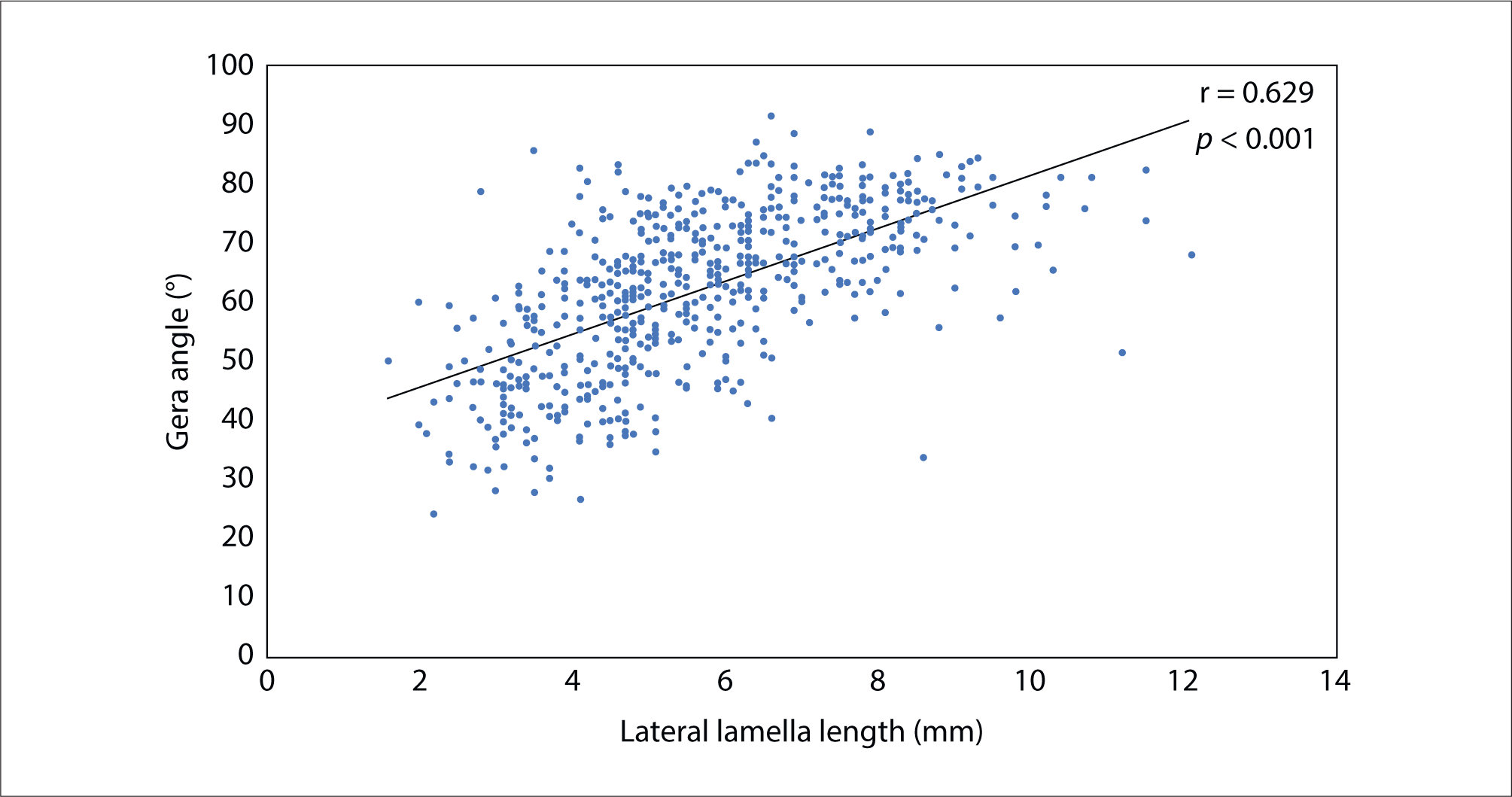
사판측벽 길이와 Gera 각도는 강한 양의 상관관계를 보였다(r=0.629, p<0.001; Fig. 2).

jcohns-35-4-145-g2
Fig. 2. Correlation between the lateral lamella length and the Gera angle.
Download Original Figure
K & G 유형

K II&G II가 가장 높은 빈도였고, K I&G I과 K III&G III가 가장 낮은 빈도를 보였다(Table 4).

Table 4. Keros & Gera combination types
Keros classification Gera classification
I II III
I 1 (0.2) 65 (11.1) 40 (6.8)
II 27 (4.6) 351 (60.1) 27 (4.6)
III 14 (2.4) 62 (10.5) 1 (0.2)

Data are presented as n (%).

Download Excel Table

고찰

후각와는 전방 두개강의 함몰부로 두개강과 비강을 분리하는데 후각와의 측면 및 중앙 경계는 사판측벽과 볏돌기이다.9)

Madhdian과 Kheir의 연구에서 Keros I, II, III형의 분포(%)는 20.43/66.26/13.31였다.5) Nitinavakarn 등10)은 11.9/68.8/19.3, Anderhuber 등11)은 14.2/70.6/15.2, Basak 등12)은 9/53/38, Ali 등13)은 20/78.7/1.3, Erdem 등14)은 8.1/59.6/32.3, Güler 등15)은 26/66/8였다. 본 연구에서는 18/68.9/13.1로 Anderhuber 등11)의 연구와 유사하였다. 연구들마다 차이는 몇 가지 이유로 추정된다. 첫째, 연구대상군의 인종 차이일 수 있다. 서양인과 동양인 등 다양한 인종간의 두부안면 부위의 해부학적 차이로 기인할 수 있다. 둘째, 부비동 CT 셋팅값 차이일 수 있다. 본 연구에서는 1 mm 두께로 축상면을 측정하였으며, 관상면과 시상면은 Wizard workstation을 통해 2 mm 두께로 재구성되었다. 셋째, 어느 관상면을 선택하는 부분에 따라 차이가 있을 수 있다. 본 연구는 관상면들 가운데 볏돌기가 잘 관찰된 부분을 선택하여 측정하였다.

Madhdian과 Kheir의 연구에서의 Gera 분류의 타입(I, II, III) 분포(%)는 29.57/61.42/9.01였다.5) Gera 등4)은 32.6/62.7/4.7, Fadda 등16)은 17.7/77.5/4.8, 본 연구에서는 7.1/81.3/11.6였다. 서양인의 경우 타입 III가 4.0%–9.0%인 반면, 본 연구에서 11.6%로 한국인에서 내시경 부비동 수술 동안 뇌기저부 손상에 더 취약할 수 있음을 시사한다.

어떤 연구에서는 남성과 여성 사이의 Keros 유형 분포에 유의한 차이를 보이지 않았으나,4,17,18) 일부 다른 연구에서는 두 성별 사이의 Keros 유형 분포가 달랐다.19,20) 본 연구에서는 두 성별 사이의 Keros 유형 분포가 달랐다(p=0.022). 본 연구에서 두 성별 사이의 Gera 타입 분포에 차이를 보이지 않았다(p=0.167). 기존 연구에서도 본 연구와 유사하게 두 성별 간의 Gera 분류에 차이를 보이지 않았다.4,6) 남성에서 여성에 비해 Keros III형의 높은 빈도는 사판측벽 길이가 평균적으로 크기 때문으로 생각되며, 일반적으로 남성에서 여성보다 큰 평균 비강 치수와 일치한다.21) 그러나 성별 간의 측정 차이가 있다고 하여 어느 성별에서 뇌 기저부 손상에 더 취약하다고 단언하기에는 무리가 있다.

본 연구에서 좌, 우측 간의 Keros 분류에서 유형의 차이를 보였으며, 양측의 사판측벽 길이 차이도 보였다(p=0.028, p<0.001). 여러 연구에서 양측 간의 사판측벽 길이의 비대칭성이 보고되었으며, 본 연구의 결과와 일치된 소견이었다.4,13,19,20,22) 본 연구에서 양측 간의 Gera 분류에서 유형 차이를 보이지 않았으나, 양측의 Gera 각도 차이는 보였다(p= 0.220, p=0.017). 두개안면 해부학 연구에서 두개골의 비대칭은 성인에서 흔히 관찰되는 소견이다.23) 사람의 약 3분의 2에서 우측보다 좌측에서 두개안면부가 보다 더 크다는 연구24)가 있어 우측 사판측벽 길이보다 좌측이 더 길었는지에 대한 추정 근거가 된다. 양측의 사판측벽 길이와 Gera 각도 차이는 내시경 부비동 수술 동안 어느 한 측이 보다 더 뇌기저부 손상에 취약할 수 있음을 시사한다.

Gera 등의 연구에서 Gera 각도와 사판의 깊이와의 유의한 양의 상관관계(r=0.553)를 보였으나, Gera 각도와 사판측벽 길이와는 유의한 음의 상관관계(r=–0.397)를 보였다. Keros 보고에서는 사판측벽 길이를 측정하여 후각와 깊이(사판측벽 길이와 후각와 깊이가 동일)를 결정한 반면,3) Gera 등의 연구에서는 Keros가 처음 제시한 측정방법과는 다르게 사판의 깊이와 사판측벽 길이를 별개로 측정하여 보고하였다.4) Gera 등의 연구에서는 사판의 깊이가 다른 연구들에서의 사판측벽 길이와 동일한 측정으로 생각된다.

Abdullah 등의 연구에서 사판측벽 길이(후각과 깊이)와 Gera 각도와의 약한 양의 상관관계(r=0.16)가 있었다.6) Madhdian과 Kheir의 연구에서는 약한 음의 상관관계(좌측: r=–0.327, 우측: r=–0.301)를 보였다.5) 본 연구에서는 유의한 양의 상관관계(r=0.629)가 있었다. 양의 상관관계는 사판측벽 길이가 길수록 Gera 각도가 커지며, 음의 상관관계는 그와는 반대이다. 즉, 유의한 음의 상관관계가 클수록 두 분류 간의 뇌 기저부의 손상위험의 일치도가 높아진다.

본 연구의 Table 4에서 고위험군인 Gera 타입 III로 분류된 68례(11.6%)의 Keros 분포 유형은 1형은 40례, II형은 27례, III형은 1례로, Keros 분류의 1형(저위험)이 III형(고위험)인 경우 보다 많아 두 분류 간의 손상위험도가 일치되지 않았다. 그리고 Keros III형(고위험)인 77례(13.1%)는 Gera 타입1이 14례, II은 62례, III은 1례로, Gera 분류의 타입 1(저위험)이 타입 III(고위험)보다 많아 두 분류 간의 손상위험도에 또한 일치된 소견을 보이지 않았다.

Preti 등은 124명의 환자를 대상으로 하여 Keros와 Gera 분류를 비교하였다.16) 그들의 연구에서 의인성 뇌척수액 유출군과 대조군 사이에 Gera 분류는 차이가 있는데 반해, Keros 분류는 유의한 차이가 없다고 보고하였다. 대조군에서는 우측의 Gera 각도가 71.7°와 좌측은 71.1°인 반면 뇌척수액 유출군에서의 유출된 측은 Gera 각도가 평균 41.2°, 반대측은 50.1°였다. 뇌척수액 유출군인 24명의 환자에서 19명이 Gera 분류에서 고위험군인 타입 III에 해당되었다. 그러나 의인성 뇌척수액 유출군의 표본수가 작기 때문에 보다 많은 연구대상군의 비교 분석이 필요할 것으로 생각된다. 그러나 적은 표본수에도 본 연구의 뇌 기저부 손상에서의 위험성 예측에서 Keros와 Gera 분류 간에 서로 상관성이 없다는 근거로 Preti 등16)의 연구 결과가 뒷받침이 될 수 있다. 본 연구를 통해 Keros와 Gera 분류 간에 서로 상관성은 없었지만, 수술 전 부비동 CT에서 Keros III형 혹은 Gera 타입 III이 관찰되는 경우 내시경 부비동 수술 동안 특히 뇌기저부의 주변 병변을 제거 시 조심스러운 접근이 필요하다.

본 연구의 강점으로 한국인을 대상으로 Gera 분류와 Keros 분류의 상관관계를 분석한 첫 연구이다. 본 연구의 제한점은 첫째, 본 연구군의 제외기준 문제이다. 제외기준으로 비부비동 병변이 영상학적 계측에 혼돈을 줄 수 있는 다발성 용종, 종양성 및 낭종성 병변, 그리고 코 수술 기왕력인데 제외기준의 경우가 실제 임상적으로 수술을 하는 경우가 많다. 둘째, 연구군의 대상 수 문제이다. 본 연구가 294명(588측)한국인 대상으로 하였으나, 더 많은 대규모의 한국인을 대상으로 한다면 환자군 선정의 선택적 오류가 최소화되리라 생각된다. 셋째, Keros와 Gera 분류에 대한 임상적 자료가 포함되어 있지 않다. 내시경 부비동 수술을 받은 연구군의 뇌기저부의 합병증이 있었던 군과 없었던 군에 Keros와 Gera 분류의 분석이 포함된다면 보다 완성도 있는 논문이 될 것으로 생각된다. 넷째, 연구 결과에 대한 부분이다. 본 연구를 포함해서 국외 연구들이 서로 상이한 결과를 보여 추가적인 많은 연구들이 필요하다. 다섯째, 다른 해부학적 요인들과의 연관성에 대한 부분이다. 전두오목(frontal recess)내 혹은 주변에 붕소의 존재 및 전사골동맥 위치와 같은 부비동 내시경 수술에서 해부학적 요인들과 두 분류와의 연관성에 대한 연구가 필요할 수 있다. Keros 분류에서 I형보다 III형에서 전사골동맥이 뇌 기저부에서 멀어져 아래 부위로 주행하는 경우가 보다 관찰되어 수술 동안 동맥 손상으로 인한 출혈가능성의 위험이 높아진다는 연구와 상안와 사골붕소(supraorbital ethmoid cell)의 존재가 낮은 위치의 전사골동맥과 연관된다는 연구가 있다.25,26) 그래서 한국인에서 Keros 분류뿐만 아니라 Gera 분류에서도 낮은 위치의 전사골동맥 및 상안와 사골붕소 존재와의 연관성에 대한 연구가 필요할 수 있다.

결론적으로, 성별 간에 Gera 각도 차이와 양측 간의 사골측벽 길이 및 Gera 각도 차이는 내시경 부비동 수술 혹은 뇌 기저부 수술에서 뇌 기저부의 개인 간 및 성별 차이가 있음을 수술전 부비동 CT을 통해 잘 파악해야 한다. 본 연구에서는 사판측벽 길이와 Gera 각도가 유의한 양의 상관관계를 보여, 뇌 기저부 손상위험도에서 Keros와 Gera 분류 간에 서로 상관성이 없을 것으로 판단된다.

Acknowledgements

Not applicable.

Funding Information

Not applicable.

Conflicts of Interest

No potential conflict of interest relevant to this article was reported.

Author Contribution

Conceptualization: Son SJ, Koo HB, Lee JH.

Data curation: Son SJ, Koo HB, Yoon K.

Formal analysis: Son SJ, Koo HB, Yoon K.

Methodology: Son SJ, Koo HB, Lee JH .

Writing-original draft: Son SJ, Koo HB, Lee JH.

Writing - review & editing: Son SJ, Koo HB, Yoon K, Lee JH.

Ethics Approval

Not applicable.

References

1.

Vaid S, Vaid N. Normal anatomy and anatomic variants of the paranasal sinuses on computed tomography. Neuroimaging Clin N Am 2015;25(4):527-48.

2.

Ohnishi T, Yanagisawa E. Lateral lamella of the cribriform plate: an important high-risk area in endoscopic sinus surgery. Ear Nose Throat J 1995;74(10):688-90.

3.

Keros P. On the practical value of differences in the level of the lamina cribrosa of the ethmoid. Z Laryngol Rhinol Otol 1962;41:809-13.

4.

Gera R, Mozzanica F, Karligkiotis A, Preti A, Bandi F, Gallo S, et al. Lateral lamella of the cribriform plate, a keystone landmark: proposal for a novel classification system. Rhinology 2018;56(1):65-72.

5.

Mahdian M, Kheir MK. CBCT assessment of ethmoid roof variations through Keros, Gera, and TMS classifications. Int J Otolaryngol 2022;2022(1):3708851.

6.

Abdullah B, Chew SC, Aziz ME, Shukri NM, Husain S, Joshua SW, et al. A new radiological classification for the risk assessment of anterior skull base injury in endoscopic sinus surgery. Sci Rep 2020;10(1):4600.

7.

Koo HB, Kown JH, Lee JH. Laterality of ethmoid roof in a Korean population. J Rhinol 2021;28(2):102-5.

8.

Son SJ, Koo HB, Lee JH. A study of skull base according to Gera classification in a Korean population. Korean J Otorhinolaryngol Head Neck Surg 2023;67(2):87-91.

9.

Jacob TG, Kaul JM. Morphology of the olfactory fossa: a new look. J Anat Soc India 2014;63(1):30-5.

10.

Nitinavakarn B, Thanaviratananich S, Sangsilp N. Anatomical variations of the lateral nasal wall and paranasal sinuses: a CT study for endoscopic sinus surgery (ESS) in Thai patients. J Med Assoc Thai 2005;88(6): 763-8.

11.

Anderhuber W, Walch C, Fock C. Configuration of ethmoid roof in children 0–14 years of age. Laryngo Rhino Otologie 2001;80(9):509-11.

12.

Başak S, Akdilli A, Karaman CZ, Kunt T. Assessment of some important anatomical variations and dangerous areas of the paranasal sinuses by computed tomography in children. Int J Pediatr Otolaryngol 2000;55(2): 81-9.

13.

Ali A, Kurien M, Shyamkumar NK, Selvaraj. Anterior skull base: high risk areas in endoscopic sinus surgery in chronic rhinosinusitis: a computed tomographic analysis. Indian J Otolaryngol Head Neck Surg 2005; 57(1):5-8.

14.

Erdem G, Erdem T, Miman MC, Ozturan O. A radiological anatomic study of the cribriform plate compared with constant structures. Rhinology 2004;42:225-9.

15.

Güler C, Uysal İÖ, Polat K, Şalk İ, Müderris T, Koşar Mİ. Analysis of ethmoid roof and skull base with coronal section paranasal sinus computed tomography. J Craniofac Surg 2012;23(5):1460-4.

16.

Preti A, Mozzanica F, Gera R, Gallo S, Zocchi J, Bandi F, et al. Horizontal lateral lamella as a risk factor for iatrogenic cerebrospinal fluid leak: clinical retrospective evaluation of 24 cases. Rhinology 2018;56(4):358-63.

17.

Salroo IN, Dar NH, Yousuf A, Lone KS. Computerised tomographic profile of ethmoid roof on basis of keros classification among ethnic Kashmiri’s. Int J Otorhinolaryngol Head Neck Surg 2016;2(1):1-5.

18.

Kaplanoglu H, Kaplanoglu V, Dilli A, Toprak U, Hekimoğlu B. An analysis of the anatomic variations of the paranasal sinuses and ethmoid roof using computed tomography. Eurasian J Med 2013;45(2):115-25.

19.

Babu AC, Nair MRPB, Kuriakose AM. Olfactory fossa depth: CT analysis of 1200 patients. Indian J Radiol Imaging 2018;28(04):395-400.

20.

Adeel M, Ikram M, Rajput MSA, Arain A, Khattak YJ. Asymmetry of lateral lamella of the cribriform plate: a software-based analysis of coronal computed tomography and its clinical relevance in endoscopic sinus surgery. Surg Radiol Anat 2013;35(9):843-7.

21.

Samoliński BK, Grzanka A, Gotlib T. Changes in nasal cavity dimensions in children and adults by gender and age. Laryngoscope 2007;117(8):1429-33.

22.

Costa ALF, Paixão AK, Gonçalves BC, Ogawa CM, Martinelli T, Maeda FA, et al. Cone beam computed tomography-based anatomical assessment of the olfactory fossa. Int J Dent 2019;2019(1):4134260.

23.

Trenouth MJ. Asymmetry of the human skull during fetal growth. Anat Rec 1985;211(2):205-12.

24.

Vig PS, Hewitt AB. Asymmetry of the human facial skeleton. Angle Orthod 1975;45(2):125-9.

25.

Poteet PS, Cox MD, Wang RA, Fitzgerald RT, Kanaan A. Analysis of the relationship between the location of the anterior ethmoid artery and Keros classification. Otolaryngol Head Neck Surg 2017;157(2):320-4.

26.

Li M, Sharbel DD, White B, Tadros SY, Kountakis SE. Reliability of the supraorbital ethmoid cell vs Keros classification in predicting the course of the anterior ethmoid artery. Int Forum Allergy Rhinol 2019;9(7): 821-4.