Case Report
재발성 뇌척수액 비루 및 뇌수막염을 일으킨 자발성 전두동 수막류 1례
A Case of Spontaneous Meningocele of the Frontal Sinus Causing Recurrent Cerebrospinal Fluid Rhinorrhea and Meningitis
Jin Hye Kwak
1
, Seogang Park
1, Seon Min Jung
1, Jin Hyeok Jeong
1,*
1한양대학교 의과대학 이비인후-두경부외과학교실
1Department of Otolaryngology-Head and Neck Surgery, College of Medicine, Hanyang University, Seoul, Korea
*Corresponding author: Jin Hyeok Jeong, Department of Otolaryngology-Head and Neck Surgery, College of Medicine, Hanyang University, Seoul 04763, Korea, Tel: +82-31-560-2296, Fax: +82-31-560-2179, E-mail:
ent@hanyang.ac.kr
© Copyright 2024 The Busan, Ulsan, Gyeoungnam Branch of Korean Society of Otolaryngology-Head and Neck Surgery. This is an Open-Access article distributed under the terms of the
Creative Commons Attribution Non-Commercial License (http://creativecommons.org/licenses/by-nc/4.0/) which permits
unrestricted non-commercial use, distribution, and reproduction in any
medium, provided the original work is properly cited.
Received: Apr 22, 2024; Revised: May 23, 2024; Accepted: Jun 02, 2024
Published Online: Jun 30, 2024
ABSTRACT
Meningocele is a bulging of the meninges through a defect in the skull or spine, forming a cystic structure filled with cerebrospinal fluid (CSF) and devoid of neural tissues. If left untreated, meningocele with CSF leakage can lead to potentially life-threatening complications such as recurrent meningitis. We report a case of spontaneous meningocele in the frontal sinus treated with endoscopic excision and reconstruction of the skull base. A 44-year-old male, with no previous history of trauma, presented with recurrent watery rhinorrhea from the right nasal cavity. An magnetic resonance imaging (MRI), conducted during treatment for recurrent meningitis, revealed a protruding mass in the right frontal sinus. Exploratory endoscopic sinus surgery exposed a meningocele with cerebrospinal fluid leakage. The meningocele was successfully treated with no signs of recurrence.
Keywords: Meningocele; Cerebrospinal fluid rhinorrhea; Meningitis
서론
수막류(meningocele)는 뇌와 척수를 둘러싸고 있는 수막 조직이 골 결손 부위를 통해 돌출되는 질환이며, 내부에 뇌 조직이 포함되지 않은 것이 특징이다. 이는 선천적으로 발생하거나, 외상이나 골 침식성 병변으로 인해 후천적으로 발생할 수 있다.1) 대부분 소아에서 선천적 병변으로 발견되며, 성인에게 발견되는 경우는 드물다.2,3) 발생 위치에 따라 증상이 다양하게 나타날 수 있으며, 특히 비내 수막류는 일측성 비루, 코막힘과 같은 증상이 나타나거나 생명의 위협이 될 수 있는 반복적인 뇌수막염이나 간질을 초래하기도 한다.4) 수막류의 진단을 위해서 전산화 단층 촬영(computed tomography, CT)과 자기공명영상(magnetic resonance imaging, MRI)이 사용된다.4,5)
저자들은 반복되는 뇌수막염을 주소로 내원한 44세 남성 환자의 전두동에서 발생한 자발성 수막류 1례를 경험하였으며, 이에 대한 문헌 고찰과 함께 보고하는 바이다.
증례
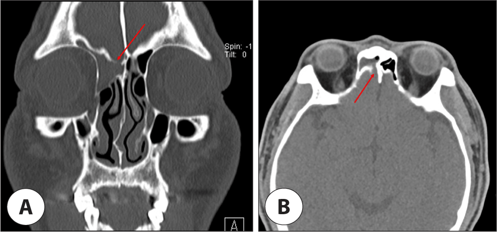
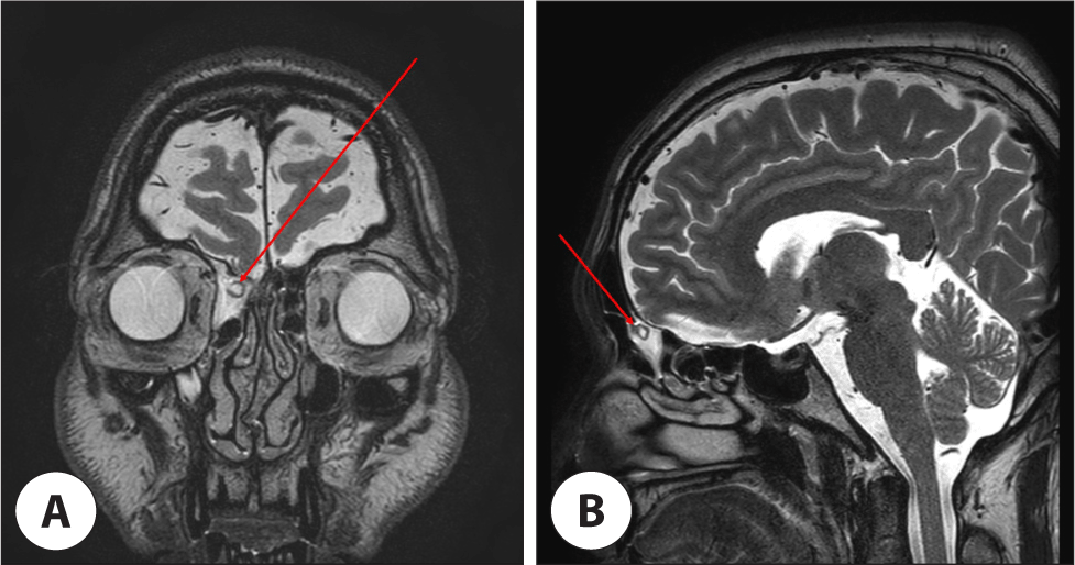
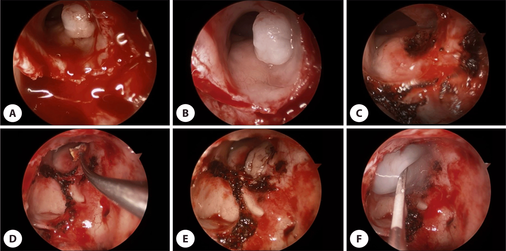
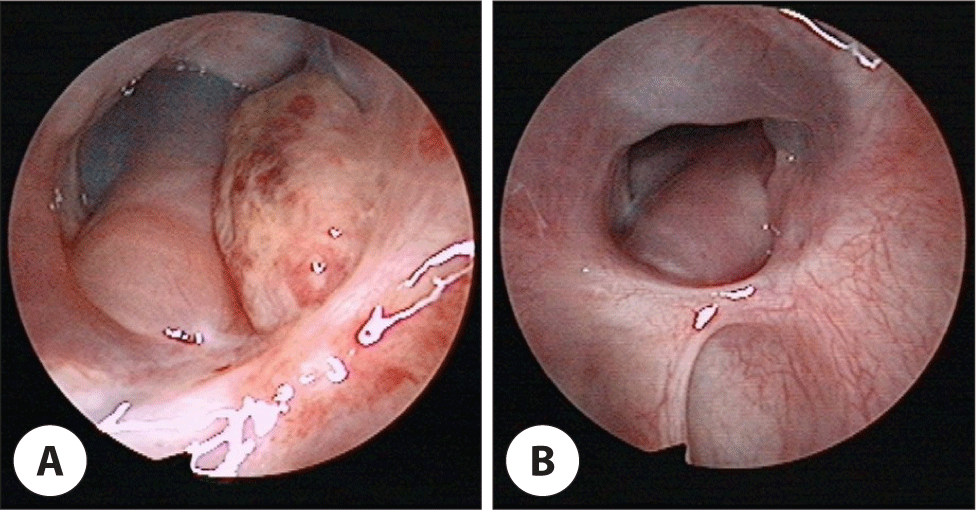
44세 남자 환자가 2주 전부터 지속된 두통으로 타 병원에서 뇌수막염 진단 하에 입원 치료를 시행하였고, 시행한 두부 영상 검사에서 우측 부비동염 의심 소견으로 본원 이비인후과로 의뢰되었다. 환자는 두통이 발생하기 2주 전부터 우측 코막힘과 수양성 비루, 오한 등의 증상이 있었으며, 2년 전에도 동일하게 뇌수막염으로 치료받은 병력이 있었다. 두부와 안면 부위의 특별한 외상력은 없었으며, 기저 질환으로는 당뇨 외 다른 질환은 없었다. 환자의 양측 비 내시경 검사상 특이 소견은 관찰되지 않았다. 시행한 부비동 CT상 우측 전두동, 전사골동, 상악동에 음영이 관찰되었고, 우측 사상판의 가측판(lateral lamella of cribriform plate)에 결손이 의심되었다(Fig. 1). 뇌 MRI에서는 우측 사상판 부위에 뇌 경질막(dura)의 결손이나 뇌실질 탈출 소견은 관찰되지 않았으며, 전두동에 뇌척수액과 동일한 신호강도를 가진 종물이 관찰되었다(Fig. 2). 환자는 수막류를 의심하고 전신마취 하 내시경 부비동 수술을 통해 수막류 제거술 및 두개저 결손 부위 재건술을 시행하였다. 구상 돌기 및 사골포를 제거한 후, 전사골동을 제거한 뒤 전두동와를 넓히자, 사상판 가측판의 골결손 부위에서 박동성으로 뇌척수액이 누출되는 하얀 낭성 종물이 발견되었다(Fig. 3A, B). 조심스럽게 종물을 제거하고(Fig. 3C), 결손 부위에 섬유소 응고제(TachocombⓇ, Nycomed, Austria)를 삽입하였다(Fig. 3D). 중비갑개 부분 절제술을 통해 채취한 중비갑개 점막을 결손 부위에 덮은 후(Fig. 3E), 트롬빈-피브리노겐 복합체(Greenplast Q, Green Cross, Cheongwon, Korea)를 도포하여 고정을 시행한 뒤(Fig. 3F), 비강을 패킹하였다. 요추배액술(lumbar drain)은 시행하지 않았고, 수술 후 침상 안정을 취하면서 예방적 항생제를 사용하였으며, 수술 후 1일째 비강 패킹을 제거하고, 4일째에 합병증 없이 퇴원하였다. 병리학적 검사 결과 섬유질 기질과 급성 및 만성 염증이 보고되었으며, 뇌실질은 관찰되지 않아 수막류에 합당한 소견이 확인되었다. 수술 후 1년의 주기적인 외래 추적 관찰에서 뇌척수액 비루가 관찰되지 않았으며, 결손 부위 이식편이 잘 이식된 채로 치유된 모습을 확인하였다(Fig. 4).
Fig. 1.
Paranasal (PNS) computed tomography (CT) images. Coronal (A) and axial (B) views of PNS CT show dehiscence (red arrow) of the right lateral lamella of the cribriform plate.
Download Original Figure
Fig. 2.
Brain magnetic resonance imaging (MRI) images. (A) Coronal view of Brain MRI T2-weighted image shows that there was a protruding mass (red 164 arrow) that looks like a meningocele and there was no involvement of brain tissue. (B) Sagittal view of Brain MRI T2-weighted image.
Download Original Figure
Fig. 3.
Operative findings. (A) A whitish mass was noted at the lateral lamella of the cribriform plate. (B) Pulsating cerebrospinal fluid (CSF) leakage was present. (C) After removing the mass. (D) Fibrin sealant patch was inserted into the defect. (E) Harvested middle turbinate mucosa was placed on the defect. (F) Fibrin glue was applied on the harvested mucosa.
Download Original Figure
Fig. 4.
Outpatient clinic follow-up. (A) 1-month after surgery. The flap is well attached at the previous cerebrospinal fluid (CSF) leakage site. (B) 9-month after surgery. The flap remains well attached with no signs of CSF leakage.
Download Original Figure
고찰
뇌류(cephalocele)는 두개골의 결손을 통해 뇌의 일부가 두개외로 돌출되어 지주막하 공간과 뇌척수액이 연결된 상태로, 이탈하는 내용물의 종류에 따라 분류된다. 뇌막만 이탈된 경우를 수막류(meningocele), 뇌실질과 뇌막이 함께 탈출하는 경우를 수막뇌류(meningoencephalocele), 뇌실까지 포함된 경우를 뇌낭수막류(encephalocystomeningocele)로 분류한다.6) 발생하는 해부학적 위치에 따라 후두형(occipital type), 전두형(sincipital type), 기저형(basal type)으로 구분된다.4) 후두형은 전체의 75%를 차지하는 가장 흔한 형태이며, 전두형은 15%, 기저형은 10%로 가장 드물게 발생한다.7) 본 증례에서는 사상판 가측판의 결손 부위에서 발생한 기저형에 해당한다. 주로 발생하는 증상으로는 지속되는 일측성 맑은 비루, 코막힘, 두통, 재발하는 뇌수막염 등이 있다.4,7)
뇌류는 선천성과 후천성으로 나뉘며, 후천성은 외상성과 자발적인 비외상성으로 세분화된다.8) 자발적으로 발생하는 경우는 드물지만, 내압의 상승과 연관될 것으로 여겨지며, 특발성 두개내 고혈압과 자발성 뇌척수액 유출은 여성과 비만인 경우에 더 높은 위험성이 보고되었다.9) 발생 기전으로는, 저항에 약한 부위에서 수막낭이 커지면서 뇌 기저부를 압박하여 골 침식을 일으키고, 수막이 비강 또는 부비동으로 탈출하며, 수막류가 천공되어 뇌척수액 비루를 일으킬 수 있다고 생각된다.8)
뇌류의 진단은 영상학적 검사를 통해 이루어진다. CT는 뇌 기저부의 결손을 확인하고 종물의 크기나 범위에 대한 정보를 제공하며, MRI는 두개 내 연결성 여부 및 수막류와 뇌수막류를 감별 진단하는 데 도움을 준다.9) 국내에는 β-2 transferrin과 β-trace 단백 검사가 없어 뇌척수액 비루의 확진이 제한적이지만, 포도당, 단백질 등 화학적 성분 분석을 통해 정보를 얻을 수 있다.10) 비강 내에서 박동성 종물이 관찰되고 수막류가 의심되는 경우, 뇌막염 등 두개 내 합병증의 위험으로 조직검사는 시행하지 않고, 방사선 검사를 통해 진단 후 수술적 제거를 시행한다.4)
돌출된 뇌류를 절제하고 뇌척수액이 누출되는 결손 부위를 재건하는 방법으로는 측면 비절개술(lateral rhinotomy), 개두술(open transcranial approach), 경비강 접근법(transnasal approach)이 있다.11–13) 종물의 크기나 위치에 따라 접근법이 달라지며, 크기가 크거나 두개 내에 있는 경우 개두술을 시행하며, 개방술만으로 결손 부위를 막기 어려울 때는 복합 접근법이 필요하다.14) 기저형 수막류의 경우, 확대된 시야의 장점과 수술 후 추적 관찰의 편리함을 비롯하여, 미용적인 측면과 수술 후 합병증 측면에서도 우수함을 제공하는 내시경하 경비강 접근법이 널리 사용되고 있다.13,15)
결론
본 증례는 성인에게서 발생한 뇌척수액 비루와 반복되는 뇌수막염을 동반한 자발성 전두동 수막류를 내시경하 경비강 접근법으로 제거하고 결손 부위를 재건하여, 재발 없이 성공적으로 치료하였기에 보고하는 바이다.