Journal of Clinical Otolaryngology Head and Neck Surgery
The Busan, Ulsan, Gyeoungnam Branch of Korean Society of Otolaryngology-Head and Neck Surgery
Original Article

비과영역과 연관된 절치공 계측에 대한 영상학적 연구

손상준1https://orcid.org/0000-0001-6403-5278, 구형본1https://orcid.org/0000-0002-1342-9450, 곽재환1https://orcid.org/0009-0008-0488-9392, 윤기서1https://orcid.org/0009-0002-4991-4041, 이재훈1,*https://orcid.org/0000-0002-0856-8173
Sang-Jun Son1https://orcid.org/0000-0001-6403-5278, Hyung-Bon Koo1https://orcid.org/0000-0002-1342-9450, Jae-Hwan Kwak1https://orcid.org/0009-0008-0488-9392, Kiseo Yoon1https://orcid.org/0009-0002-4991-4041, Jae-Hoon Lee1,*https://orcid.org/0000-0002-0856-8173
1원광대학교 의과대학 이비인후과학교실
1Department of Otolaryngology-Head and Neck Surgery, Wonkwang University School of Medicine, Iksan, Korea
*Corresponding author: Jae-Hoon Lee, Department of Otolaryngology-Head and Neck Surgery, Wonkwang University School of Medicine, Iksan 54538, Korea, Tel: +82-63-859-1441, Fax: +82-63-841-6556, E-mail: Leejaehoon64@gmail.com

© Copyright 2024 The Busan, Ulsan, Gyeoungnam Branch of Korean Society of Otolaryngology-Head and Neck Surgery. This is an Open-Access article distributed under the terms of the Creative Commons Attribution Non-Commercial License (http://creativecommons.org/licenses/by-nc/4.0/) which permits unrestricted non-commercial use, distribution, and reproduction in any medium, provided the original work is properly cited.

Received: Apr 11, 2024; Revised: May 15, 2024; Accepted: Jun 13, 2024

Published Online: Jun 30, 2024

ABSTRACT

Background and Objectives:

Most episodes of the anterior epistaxis are controlled by the anterior pressure methods. If the pressure methods fail, surgical interventions such as vessel cauterization, ligation, and embolization can be recommended. While surgical techniques for the sphenopalatine artery and anterior ethmoidal artery are well-described, the technique for the greater palatine artery (GPA) is not as well-known. The purpose of this study was to describe the incisive foramen, the exit of the GPA, and the surrounding structures using sinus computed tomography (CT).

Materials and Methods:

Sinus CT was performed on a total of 233 individuals. Using sagittal images, the distance from the anterior nasal spine to the incisive foramen (DANSIF) and the antero-posterior diameter of the incisive foramen (DIF) were measured.

Results:

The DANSIF in males (11.63±2.44 mm) was longer than that in females (10.62±2.19 mm), showing significant gender difference (p=0.001). The DIF in males (4.43±1.43 mm) was longer than that in females (4.21±1.34 mm), showing no significant gender difference (p=0.244). The DANSIF and age were negatively correlated (r=–0.318, p<0.001).

Conclusion:

In Korean adults, the DANSIF and DIF are slightly longer than 10 mm and 4 mm, respectively. Measuring the DANSIF is a useful method for finding the exit of the GPA in the nasal cavity.

Keywords: Epistaxis; Greater palatine artery; Anterior nasal spine; Incisive foramen; Computed tomography

서론

비출혈의 약 80%는 상순동맥(superior labial artery), 대구개동맥(greater palatine artery), 접형구개동맥(sphenopalatine artery) 및 전사골동맥(anterior ethmoidal artery)의 분지들이 합류하는 리틀영역(Little’s area)에서 발생한다.1,2) 대부분 비출혈은 보존적인 치료로 조절된다. 팩킹, 소작 등의 치료로 잘 조절되지 않는다면, 원인 의심이 되는 혈관에 대한 직접 소작/결찰 및 색전술과 같은 보다 적극적인 치료 방법이 선택될 수 있다.37) 접형구개동맥과 전사골동맥에 대한 혈관소작 및 결찰에 대한 술기는 잘 알려져 있다.3,5,8)

대구개동맥의 주요 줄기는 절치공(incisive foramen)을 통해 비강으로 들어가는데, 비중격의 전하부에서 접형구개동맥과 전사골동맥의 중격 분지들과 접합한다.9) 그러나 적절한 치료에도 불구하고 혈관 확장증(vascular ectasia)과 같은 경우 빈번하게 전방 출혈이 발생되어 고통이 심하게 되므로 혈관에 대해 직접적인 소작 혹은 결찰이 필요할 수 있다.5)

대구개동맥은 보존적인 치료에 반응하지 않는 빈번한 전방 출혈의 원인이 될 수 있으며, 대구개동맥 유경피판(pedicled flap)을 이용한 비중격 천공 재건에 이용될 수 있다.10,11) 또한, 구강상악동루(oroantral fistula) 재건에 대구개동맥 유경피판이 사용되었다.12)

대구개동맥의 비과적인 임상적 활용을 위해서는 전비극에서 절치공까지의 거리 측정을 통한 대구개동맥의 위치파악이 중요하다. 아직까지 한국인을 대상으로 한 연구는 없었다. 그래서 저자들은 부비동 컴퓨터 단층 촬영(computed tomography, CT)을 이용하여 대구개동맥의 비강으로의 출구인 절치공의 해부학적 위치 및 직경에 대해 파악하고, 성별 및 나이에 따라 차이점이 있는지 알아보기 위해 본 연구를 수행하였다.

방법

부비동 CT는 Somatom Definition Flash 256-slice CT scanners(Simens Healthineers, Forchheim, Germany)를 이용하여 1 mm 두께로 축상면(axial)을 측정하였으며, 관상면(coronal)과 시상면(sagittal)은 Wizard workstation(Simens Healthineers)을 통해 2 mm 두께로 재구성되었다. 본 연구는 본 병원 기관심의위원회의 승인(2023-07-029)을 받고 수행되었다.

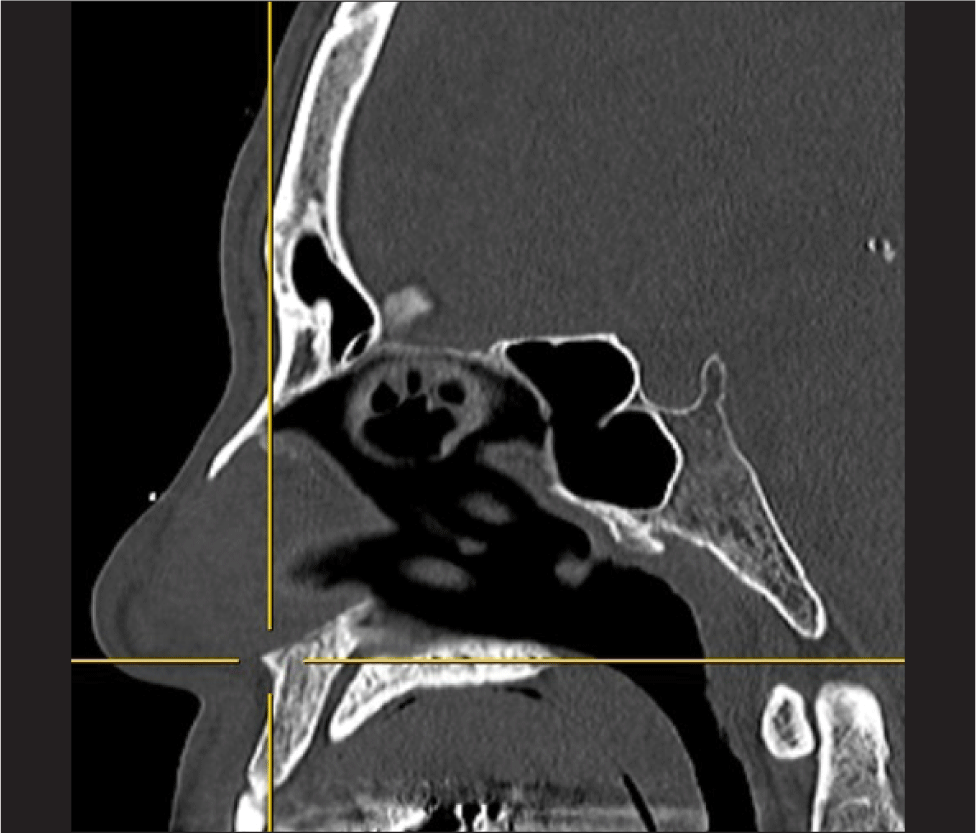
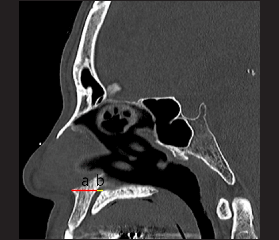
본 연구는 부비동 CT의 시상면 영상에서 전비극(anterior nasal spine)이 시작되는 부위에 3차원 커서를 적용한 후(Fig. 1), 전비극으로부터 절치공의 전단 경계까지의 수평 거리와 전비극과 절치공의 전단 경계의 연장선 상에 있는 절치공의 전후 직경을 측정하였다(Fig. 2).
jcohns-35-2-75-g1
Fig. 1. Sagittal sinus computed tomography (CT) image shows the anterior nasal spine and incisive foramen using a three-dimensional cursor.
Download Original Figure
jcohns-35-2-75-g2
Fig. 2. A sagittal image shows (a) the measurement between the anterior nasal spine and incisive foramen, and (b) the antero-posterior diameter of incisive foramen. The a (red line) and b (yellowish line) were 14.26 mm and 5.12 mm, respectively.
Download Original Figure
통계

통계학적 분석은 SPSS version 24.0 program(IBM, Armonk, NY, USA)을 이용하였다. 남녀 간에서 연속성 변수와의 분석은 Student t-검정을 사용하였다. 연령과 연속성 변수와의 분석은 Pearson 상관관계 분석(Pearson, London, UK)을 사용하였다. 통계학적 유의성은 p값이 0.05 미만인 경우에 통계적으로 유의하다고 판정하였다.

결과

연구대상군 특성(Table 1)

2021년 3월부터 2022년 3월까지 본 병원에 비과적 증상으로 내원하여 부비동 CT을 촬영했던 총 287명을 대상으로 연구를 수행하였다. 연구대상군의 제외는 선천성 구강악안면기형이 있는 2명, 안면 골절 기왕력 혹은 수술력(안면 또는 비중격 교정술 혹은 재건술)이 있는 15명, 20세 미만인 37명이었다.

Table 1. Demographics of the study group
Characteristic Value
Sex
 Male: Female 138(59.2): 95(40.8)
 Age (years) 46.7±14.6: 48.2±15.3
ANS-IF (distance)
 Mean 11.22±2.39
 Minimum 4.27
 Maximum 19.16
IF (A-P diameter)
 Mean 4.34±1.40
 Minimum 1.57
 Maximum 8.75

Data are presented as n (%) or mean±SD (mm).

ANS-IF: anterior nasal spine-incisive foramen, A-P: antero-posterior.

Download Excel Table

총 233명중 남자는 138명, 여자는 95명으로, 남자는 46.7±14.6세, 여자는 48.2±15.3세로 성별 간의 유의한 차이는 없었다(p=0.352). 전비극에서 절치공까지 거리의 최솟값은 4.27 mm, 최댓값은 19.16 mm, 평균은 11.22±2.39 mm였다. 절치공의 전후 직경의 최솟값은 1.57 mm, 최댓값은 8.75 mm 평균은 4.34±1.40 mm였다.

전비극에서 절치공까지 거리(Table 2)

연구대상군 233명 중 138명 남자의 평균은 11.63±2.44 mm, 95명 여자의 평균은 10.62±2.19 mm로 성별 간에 유의한 차이가 있었다(p=0.001). 남녀의 평균은 11.22±2.39 mm였다.

Table 2. Parameters for incisive foramen
Value Male Female p-value
ANS-IF (distance) 11.63±2.44 10.62±2.19 0.001
 Total 11.22±2.39
IF (A-P diameter) 4.43±1.43 4.21±1.34 0.244
 Total 4.34±1.40

Data are mean±SD (mm).

ANS-IF: anterior nasal spine-incisive foramen, A-P: antero-posterior.

Download Excel Table
절치공의 전후 직경(Table 2)

연구대상군 233명 중 138명 남자의 평균은 4.43±1.43 mm, 95명 여자의 평균은 4.21±1.34 mm로 성별 간에 유의한 차이를 보이지 않았다(p=0.244). 남녀의 평균은 4.34±1.40 mm였다.

전비극에서 절치공까지 거리와 연령과의 관계

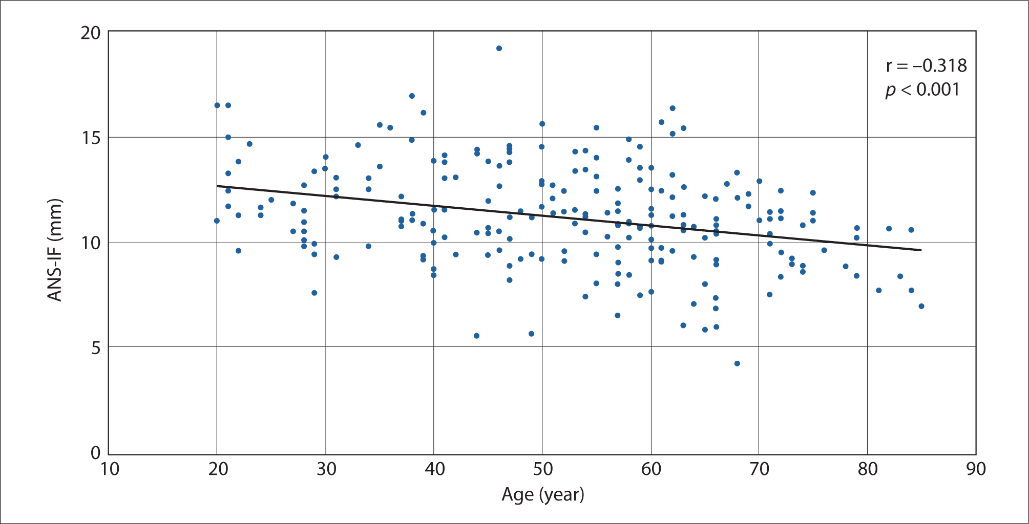
음적인 상관관계를 보였다(r=–0.318, p<0.001)(Fig. 3).
jcohns-35-2-75-g3
Fig. 3. Correlation the measurement between the anterior nasal spine and incisive foramen, and age (r=–0.318, p<0.001). ANS-IF: anterior nasal spine-incisive foramen.
Download Original Figure

고찰

절치공의 위치의 임상적 적용은 첫째, 사전에 촬영된 부비동 CT의 시상면에서 전비극에서 절치공까지의 거리를 측정한다. 둘째, 소독된 수술자에 측정된 전비극에서 절치공까지의 거리를 표시한다. 셋째, 비강 내의 위치는 촉진을 통해 전비극을 확인한 후 전비극을 기준으로 절치공까지의 거리를 소독된 수술자를 비강 내로 삽입하여 절치공의 예상되는 위치에 수술용 마커로 착색한다. 따라서, 전비극은 절치공의 위치 파악에 중요한 랜드마크로 여겨진다.

대구개동맥의 소작에 대한 접근 방법은 비중격 교정술과 유사하며 점막 박리는 절치공 부위의 뒤쪽, 아래쪽으로 진행해야 하며, 비중격과 비강 바닥 사이의 경계에서 대구개동맥이 나오므로 주의 깊은 해부학적 접근이 필요하다. 절치공에서 대구개동맥이 확인되면 바늘 끝 소작기(needle-point cautery)를 적절히 구부려서 절치공 내의 대구개동맥을 소작한다.

대구개동맥 접근으로 인해 발생 가능한 합병증은 첫째, 비중격 혈종 혹은 천공이고 둘째, 대구개동맥에 대한 직접 소작으로 인해 경구개와 중앙 앞니에 대한 감각 이상이다.5) 그 이유로 비구개신경(nasopalatine nerve)이 절치공에서 대구개동맥과 같이 위치하고 있어 대구개 동맥에 대한 소작하는 과정에서 손상을 받을 수 있다. 비구개 신경은 경구개와 중앙 앞니(central incisor)에 대한 감각을 담당한다.

Santamaría-Gadea 등의 연구에서 절치공의 영상학적 계측은 전비극에서 15±2.0 mm였다.11) Alobid 등은 비중격 전방 천공의 재건에 대해 대구개동맥 유경피판을 이용한 증례를 보고하였다.9) 수술 전 시행했던 부비동 CT의 시상면에서 절치공을 측정한 후, 그 지점을 비강 내에 전비극을 기준으로 15 mm 떨어진 곳에 표시하였다. 그 다음으로 대구개동맥을 기반으로 한 유경피판을 사용한 후 재건에 성공하였다. Tȍzȕm 등은 절치관(incisive canal)의 상부, 중간, 하부위치에서 절치관의 앞쪽에 있는 뼈의 두께에 대해 영상학적 계측을 하였는데, 각각 평균이 9.19±2.48 mm, 6.68±1.57 mm, 5.62±1.56 mm였다.13) 절치관의 상부위치에서의 절치관 앞쪽에 있는 뼈의 두께가 본 연구에서의 전비극에서 절치공까지 거리에 해당된다. 본 연구에서 전비극에서 절치공까지의 거리는 남자 평균은 11.63±2.44 mm, 여자는 10.62±2.19 mm로 전체적으로 11.22±2.39 mm였다. 연구자들 사이의 측정값 차이는 각각 개인 차이뿐만 아니라 인종간의 얼굴 모양에 따라 영향을 받을 수 있으며,1416) 연구자들 사이에서의 측정방법 차이에서 발생될 수 있다.

본 연구에서 절치공의 전후 직경은 남자 평균은 4.43±1.43 mm, 여자는 4.21±1.34 mm로 전체적으로 4.34±1.40 mm였다. 절치공의 전후 직경의 최소값은 1.57 mm, 최대값은 8.75 mm으로 개인에 따라 차이가 컸다. Tȍzȕm 등의 연구에서 절치관의 상부, 중간, 하부위치에서 전후 직경을 측정하였는데, 각각 평균이 2.76±1.40 mm, 2.07±0.92 mm, 2.93±1.01 mm였다.13) 상부위치에 대한 평균인 2.76±1.40 mm와 비교하면 본 연구에서의 측정값이 더 컸다. Soumya 등은 절치관의 중간위치에서 전후 직경과 너비를 측정하였는데, 각각 평균이 3.12±0.94 mm, 3.23±0.98 mm였다.17) 절치관의 직경에 대해 한국인을 대상으로 한 연구에서 직경의 범위는 1.5–6.4 mm였으며, 상부에 위치한 비강측 절치관에서 하부에 위치한 구개측 절치관으로 주행할수록 직경이 좁아진다고 하였다.18) 한국인을 대상으로 한 다른 연구에서 비강측 절치관의 직경은 평균 5.3 mm, 구개측 절치관의 직경은 평균 4 mm로 비강측에서 넓어졌다.19) 문헌 고찰에서 절치공 직경의 크기에 따른 임상적 의의에 대해 찾아볼 수는 없었으나, 본 저자들은 대구개동맥에 대한 전기적 소작하는 과정에서 절치공 직경의 크기 차이가 비구개 신경 손상에 영향을 주는 한 가지 요인으로 추측하였다. Bodin 등은 절치공의 직경이 6–10 mm인 경우에는 낭성 변형의 가능성이 있으므로 직경의 증가 여부를 반드시 추적 관찰해야 한다고 하였다.20)

본 연구에서 연령과 전비극에서 절치공까지 거리와의 상관관계 분석에서 음적인 상관 관계를 보였는데, 이는 노화에 따른 안면골의 리모델링과 연관성이 있어 측정거리가 감소되었을 것으로 보인다.21) Soumya 등은 절치관의 중간위치에서 절치관의 앞쪽에 있는 뼈의 두께를 30대 이하, 31세 이상 50세까지, 51세 이상의 세 군으로 나누어 평균을 구한 결과는 각각 6.73±1.67 mm, 6.3±1.33 mm, 6.02±1.34 mm로 통계학적 차이를 보이지 않았으나 연령의 증가에 따른 뼈의 두께가 감소하는 경향을 보였는데,17) 본 연구 결과와 유사한 소견이었다.

비중격천공의 원인으로는 의인성(비강 수술), 외상, 코후비기, 코이물, 전신 염증성 질환 또는 화학 물질 노출 등으로 다양하다.9) 내시경의 도입과 발달은 비중격천공의 재건에도 사용되었는데 내시경하에 전사골동맥 유경피판을 이용한 재건은 성공률이 높은 편이다.22,23) 비중격 전방의 천공부위는 전사골동맥 유경에 비해 대구개동맥 유경이 근접하고 있어서, 대구개동맥 유경피판을 사용하면 천공을 보다 쉽게 재건할 수 있다.11) 대구개동맥 유경피판을 만드는데 전비극에서 절개공까지의 거리 측정은 대구개동맥을 찾는데 유용하다. 대구개동맥 유경피판술은 절치공 출구 0.5 cm 전방의 천공에 적용될 수 있다.911)

구강상악동루는 상부 소구치 혹은 대구치 발치, 치과 감염, 외상, 방사선 치료, 골수염, 또는 치과 임플란트로 인한 구강과 상악동 사이의 연결된 통로이다.2426) 국소 및 연조직 피판, 이식(graft), 무생체소재(alloplastic materials), 금속을 포함하여 많은 기술이 구강상악동루 재건에 사용되었다.24) 비강에서 대구개동맥 유경피판 및 구강에서 협측 지방 패드 피판(buccal fat pad flap)와 같은 이중 레이어 피판을 사용하면 구강상악동루의 재건에 효과적이다.12)

본 연구의 강점으로 한국인을 대상으로 절치공의 위치를 부비동 CT에서 처음 계측한 연구로, 233명을 대상으로 한 비교적 대규모 연구이다. 부비동 CT의 시상면 영상에서 전비극에서 절치공까지의 거리 측정을 한 후 수술 시야에서 이비인후과 의사에게 익숙한 비중격 수술 접근 방법을 통해 부비동 CT의 계측치를 쉽게 적용할 수 있다. 본 연구의 제한점은 본 연구에서는 해부학적 지표를 전비극을 기준으로 절치공까지의 거리와 절치공의 전후 직경을 측정하였는데, 절치공의 다른 해부학적 지표를 통한 측정이 절치공에 대한 해부학적 이해에 보다 도움이 될 수 있을 것으로 생각된다. 후비극(posterior nasal spine)에서 절치공까지의 거리, 절치공의 너비, 절치관의 길이 등과 같은 추가적인 측정이 해당될 수 있다.

결론적으로 연구대상군의 전비극에서 절치공까지 거리와 절치공의 전후 직경은 각각 평균 10 mm, 4 mm보다 약간 컸다. 부비동 CT를 이용한 전비극에서 절치공까지의 거리 측정은 대구개동맥의 비강 출구 위치를 확인하는데 유용한 방법이다.

Acknowledgements

Not applicable.

Funding Information

This paper was supported by Wonkwang University in 2024.

Conflicts of Interest

No potential conflict of interest relevant to this article was reported.

Author Contribution

Conceptualization: Son SJ, Koo HB, Lee JH.

Data curation: Son SJ, Koo HB, Kwak JH, Yoon K.

Formal analysis: Son SJ, Koo HB, Kwak JH, Yoon K.

Methodology: Son SJ, Koo HB, Lee JH.

Writing - original draft: Son SJ, Koo HB, Lee JH.

Writing - review & editing: Son SJ, Koo HB, Kwak JH. Yoon K, Lee JH.

Ethics Approval

This study was conducted with approval from the Institutional Review Board of Wonkwang University Hospital (2023-07-029).

References

1.

Schaitkin B, Strauss M, Houck JR. Epistaxis: medical versus surgical therapy: a comparison of efficacy, complications, and economic considerations. Laryngoscope 1987;97(12):1392-6.

2.

Janfaza P, Nadol JB, Galla RJ, Fabian RL, Montgomery WW. Surgical anatomy of the head and neck. 1st ed. Philadelphia, PA: Lippincott, Williams & Wilkins; 2001. p. 283-4.

3.

Woolford TJ, Jones NS. Endoscopic ligation of anterior ethmoidal artery in treatment of epistaxis. J Laryngol Otol 2000;114(11):858-60.

4.

Dubel GJ, Ahn SH, Soares GM. Transcatheter embolization in the management of epistaxis. Semin Intervent Radiol 2013;30(3):249-62.

5.

Butrymowicz A, Weisstuch A, Zhao A, Agarwal J, Pinheiro-Neto CD. Endoscopic endonasal greater palatine artery cauterization at the incisive foramen for control of anterior epistaxis. Laryngoscope 2016;126(5):1033-8.

6.

Moreau S, De Rugy MG, Babin E, Courtheoux P, Valdazo A. Supraselective embolization in intractable epistaxis: review of 45 cases. Laryngoscope 1998; 108(6):887-8.

7.

Bradley Strong E, Antonio Bell D, Johnson LP, Jacobs JM. Intractable epistaxis: transantral ligation vs. embolization: efficacy review and cost analysis. Otolaryngol Head Neck Surg 1995;113(6):674-8.

8.

Rudmik L, Smith TL. Management of intractable spontaneous epistaxis. Am J Rhinol Allergy 2012;26(1):55-60.

9.

Alobid I, Santamaría-Gadea A, Mariño-Sánchez F. Endoscopic “racket-on-donut” technique for large anterior nasoseptal perforations. Laryngoscope 2023;134(1): 143-7.

10.

Mariño-Sánchez F, Santamaría-Gadea A, Vaca M. Technique to repair a septal perforation endoscopically with a greater palatine artery pedicled flap. Facial Plast Surg Aesthet Med 2020;22(4):301-3.

11.

Santamaría-Gadea A, Vaca M, de Los Santos G, Alobid I, Mariño-Sánchez F. Greater palatine artery pedicled flap for nasal septal perforation repair: radiological study and case series. Eur Arch Otorhinolaryngol 2021; 278(6):2115-21.

12.

Arana-Fernández B, Santamaría-Gadea A, Almeida-Parra F, Mariño-Sánchez F. Oroantral fistula closure using double-layered flap: greater palatine artery flap and buccal fat pad. Laryngoscope 2023;133(8):1824-7.

13.

Tözüm TF, Göncü GN, Yıldırım YD, Yılmaz HG, Galindo-Moreno P, Velasco-Torres M, et al. Evaluation of maxillary incisive canal characteristics related to dental implant treatment with computerized tomography: a clinical multicenter study. J Periodontol 2012; 83(3):337-43.

14.

Hwang HS, Kim WS, McNamara JA Jr. Ethnic differences in the soft tissue profile of Korean and European-American adults with normal occlusions and well-balanced faces. Angle Orthod 2002;72(1):72-80.

15.

Wen YF, Wong HM, Lin R, Yin G, McGrath C. Inter-ethnic/racial facial variations: a systematic review and bayesian meta-analysis of photogrammetric studies. PLOS ONE 2015;10(8):e0134525.

16.

Rhee SC. Differences between Caucasian and Asian attractive faces. Skin Res Technol 2018;24(1):73-9.

17.

Soumya P, Koppolu P, Pathakota KR, Chappidi V. Maxillary incisive canal characteristics: a radiographic study using cone beam computerized tomography. Radiol Res Pract 2019;2019(1):6151253.

18.

Kim GT, Hwang EH, Lee SR. A study of incisive canal using a cone beam computed tomography. Korean J Oral Maxillofac Radiol 2004;34(1):7-12.

19.

Kim SJ, Lim SH. Anatomic study of the incisive canal in relation to midpalatal placement of mini-implant. Korean J Orthod 2009;39(3):146-58.

20.

Bodin I, Isacsson G, Julin P. Cysts of the nasopalatine duct. Int J Oral Maxillofac Surg 1986;15(6):696-706.

21.

Pessa JE, Desvigne LD, Zadoo VP. The effect of skeletal remodeling on the nasal profile: considerations for rhinoplasty in the older patient. Aesthetic Plast Surg 1999;23(4):239-42.

22.

Castelnuovo P, Ferreli F, Khodaei I, Palma P. Anterior ethmoidal artery septal flap for the management of septal perforation. Arch Facial Plast Surg 2011;13(6):411-4.

23.

Santamaría-Gadea A, Langdon C, Alobid I. Extended anterior ethmoidal artery flap: novel endoscopic technique for large septal perforation. Laryngoscope 2022;132(2):298-300.

24.

Parvini P, Obreja K, Begic A, Schwarz F, Becker J, Sader R, et al. Decision-making in closure of oroantral communication and fistula. Int J Implant Dent 2019; 5(1):13.

25.

Kim MK, Han W, Kim SG. The use of the buccal fat pad flap for oral reconstruction. Maxillofac Plast Reconstr Surg 2017;39(1):5.

26.

Candamourty R, Jain MK, Sankar K, Ramesh Babu MR. Double-layered closure of oroantral fistula using buccal fat pad and buccal advancement flap. J Nat Sci Biol Med 2012;3(2):203-5.