Journal of Clinical Otolaryngology Head and Neck Surgery
The Busan, Ulsan, Gyeoungnam Branch of Korean Society of Otolaryngology-Head and Neck Surgery
Original Article

한국인에서 전두동 자연공의 위치에 따른 등급체계에 대한 영상학적 연구

구형본1https://orcid.org/0000-0002-1342-9450, 곽재환1https://orcid.org/0009-0008-0488-9392, 이재훈1,*https://orcid.org/0000-0002-0856-8173
Hyung-Bon Koo1https://orcid.org/0000-0002-1342-9450, Jae-Hwan Kwak1https://orcid.org/0009-0008-0488-9392, Jae-Hoon Lee1,*https://orcid.org/0000-0002-0856-8173
1원광대학교 의과대학 이비인후과학교실
1Department of Otolaryngology-Head and Neck Surgery, Wonkwang University School of Medicine, Iksan, Korea
*Corresponding author: Jae-Hoon Lee, Department of Otolaryngology-Head and Neck Surgery, Wonkwang University School of Medicine, Iksan 54538, Korea, Tel: +82-63-859-1441, Fax: +82-63-841-6556, E-mail: Leejaehoon64@gmail.com

© Copyright 2024 The Busan, Ulsan, Gyeoungnam Branch of Korean Society of Otolaryngology-Head and Neck Surgery. This is an Open-Access article distributed under the terms of the Creative Commons Attribution Non-Commercial License (http://creativecommons.org/licenses/by-nc/4.0/) which permits unrestricted non-commercial use, distribution, and reproduction in any medium, provided the original work is properly cited.

Received: Oct 31, 2023; Revised: Dec 11, 2023; Accepted: Jan 05, 2024

Published Online: Jun 30, 2024

ABSTRACT

Background and Objectives:

Endoscopic frontal sinus surgery (EFSS) is much more challenging than endoscopic surgery on the other paranasal sinuses. Recently, a new computed tomography (CT) grading system according to the anatomical position of the frontal ostium was introduced. The purpose of this study is to apply the new CT grading system in Koreans.

Materials and Methods:

Sinus CT was performed on a total of 233 people, and a study was conducted on bilateral 466 sides. In this study, the parameters were included 1) frontal ostium grade (FOG): positive, neutral, and negative, 2) frontal ostium diameter (FOD), 3) four levels (I–IV) based on the FOG and FOD.

Results:

Overall, the FOG was 386 (82.8%) positive, 61 (13.1%) neutral, and 19 (4.1%) negative and the average FOD was 9.43±2.80 mm. When comparing men and women, there was no significant difference in the frequency of FOG, but in the mean of FOD, men were significantly longer than women. Overall, level I was 190 sides (40.8%), level II was 224 sides (48.1%), level III was 49 sides (10.5%), and level IV was 3 sides (0.6%). When comparing men and women, there was a significant difference in the frequency of levels.

Conclusion:

This is the first study to apply the new classification based on the FOG in Koreans. However, due to the limitations of this study, there is insufficient evidence to relate to the difficulty of EFSS because the actual operation time and operation success rate according to the new classification based on FOG were not evaluated. In the future, it is thought that further clinical studies based on the results of this study should be conducted.

Keywords: Frontal sinus; Grade; Ostium; Position; Computed tomography

서론

내시경 전두동 수술은 다른 부비동 내시경 수술보다 어려움이 있는데, 두개나 안와 등의 중요 구조물에 대한 합병증이 발생할 가능성이 있으며, 복잡 다양하고 좁은 해부학적 구조를 가지고 있기 때문이다.1-3) 이러한 해부학적 구조는 전두오목(frontal recess) 내 혹은 주변에 다양한 봉소들이 존재하고, 전두동 자연공의 위치와 직경에서 변이를 보이는 경우가 많다. 전두오목은 전두오목 주변의 다양한 봉소들의 존재로 인해 전두오목이 폐쇄 혹은 좁아지고, 구상돌기의 상부 부착부위와 같은 해부학적 요인들이 관여될 수 있다.

전두오목 주변의 다양한 봉소들의 분류는 1996년 Kuhn 등에 의해 체계화되었다.1) Kuhn 등은 전두봉소(frontal cells)를 1형에서 4형까지의 4가지로 구분하였고, 2016년에 Wormald 등4)은 International Frontal Sinus Anatomy Classification(IFAC) 분류를 발표하였다. Kuhn 분류는 전두동 유출로와 관련된 주변 해부학적 구조와의 관계에 초점을 맞춘 반면, IFAC는 봉소들의 위치를 전방, 후방, 내측으로 구분하여 보다 더 수술적 접근에 초점을 맞춘 분류라 할 수 있다. 국내에서도 Kuhn 분류에 따른 연구들이 진행되어 왔다.5,6)

전두오목 주변의 다양한 봉소들에 대한 많은 관심에 비해, 전두동 자연공의 위치에 대해서는 상대적으로 관심이 적었다. Hilger 등7)은 시상면에서 전두동이 보다 앞에 위치할수록 전두-비강 각도(fronto-nasal-angle)가 작아지고, 전두동이 보다 후방에 위치할수록 전두-비강 각도가 커진다. 전두-비강 각도가 작을수록 전두동 자연공의 시야 및 접근을 위해서는 더 각도가 있는 내시경이 필요하다고 하였다. DelGaudio 등8)은 전두동 자연공은 시상면에서 가장 뒤쪽으로 돌출된 코 부리(nasal beak)에서 전두동의 가장 후벽(전방 전사골동 천장과 만나는 지점)까지의 거리(직경)로 측정하였다.

Gheriani 등9)은 부비동 단층촬영(computed tomography, CT)을 이용하여 전두동 자연공의 해부학적 위치와 직경에 따라 새로운 분류 체계를 발표하였다. 전두동 자연공의 위치를 3단계로, 전두동 자연공 직경의 중앙값을 기준으로 2분류로 구분하였다.

본 연구에서는 대규모의 한국인을 대상으로 하여 영상 해부학적 측정을 통해 Gheriani 등9)이 새롭게 제시한 전두동 자연공의 위치에 따른 분류를 하고자 하였다.

대상 및 방법

부비동 CT는 Somatom Definition Flash 256-slice CT scanners(Simens Healthineers, Forchheim, Germany)를 이용하여 1 mm 두께로 축상면을 측정하였으며, 관상면과 시상면은 Wizard workstation(Simens Healthcare)을 통해 2 mm 두께로 재구성되었다. 본 연구는 본 병원 기관심의위원회의 승인(2023-07-023)을 받고 수행되었다.

본 연구는 Gheriani 등9)의 방법을 참고하였다. 관상면에서 좌우측 각각 누낭(lacrimal sac)의 가장 내측에 3차원 커서를 적용하여 그 지점에 해당되는 시상면을 확인하였다. 그 시상면 영상과 전, 후의 시상면 영상 2장을 포함한 총 3장에서 상악골의 전두 돌기(frontal process of maxilla)의 수직 축을 따라 R선(reference line)이라고 불리는 첫 번째 선을 그었으며, 이 수직선은 코 부리(nasal beak)의 가장 뒤쪽 돌출부를 지나게 된다. S선(secondary line)은 같은 시상면에서 뇌 기저부에서 전두동 후면으로의 상승점(point of upturn)을 통과하는 수직선을 긋는데 이 수직선은 R선과 평행하게 긋는다.

전두동 단계(frontal ostium grade, FOG)는 R선이 S선의 앞쪽에 있는 경우를 양성(positive), R선과 S선이 겹치는 경우는 중성(neutral), 만약 R선이 S선의 뒤쪽에 있다면 음성(negative)으로 분류하였다(Fig. 1). Gheriani 등9)의 방법에 따라 전두동 수술의 난이도 순서는 양성(positive)이 가장 낮아 접근이 가장 용이하며, 그 다음으로 중성(neutral), 가장 높은 경우는 음성(negative)이었다.

jcohns-35-2-69-g1
Fig. 1. Frontal ostium grading. R line presents a vertical axis of the most posterior projection of the nasal beak. S line presents a vertical axis of the posterior wall of the frontal sinus (corresponding point of upturn of the anterior skull base). If the R line was ahead of the S line, we categorized frontal ositum grade as positive (A), if the R line overlapped the S line, the frontal ostium grade as neutral (B), and if the R line was posterior to the S line, the frontal ostium grade as negative (C). Red lines present frontal ostium diameter.
Download Original Figure

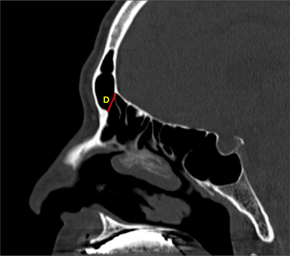
R선이 코 부리와 교차하는 지점과 S선이 뇌 기저부와 교차하는 지점 사이에 그려진 선은 세번째 선(third line [diameter: D])으로 하였다(Fig. 2). FOG의 양성 혹은 음성을 포함한 어떠한 결과값에 관계없이 이 선의 거리를 전두동 자연공의 직경(frontal ostium diameter, FOD)으로 하였다. 본 연구에서는 한 측당 측정된 3개의 시상면 영상들 중 FOD가 가장 큰 영상에서의 FOG 단계와 FOD을 선택하였다. 본 연구 대상인 466측의 FOD의 중앙값(median value)은 9.14 mm였으며, FOG와 FOD(median)를 기반으로 레벨 I은 FOG(양성)와 FOD(≥9.14 mm), 레벨 II는 FOG(중성)와 FOD(≥9.14 mm) 혹은 FOG(양성)와 FOD(<9.14 mm), 레벨 III는 FOG(음성)와 FOD(≥9.14 mm) 혹은 FOG(중성)와 FOD(<9.14 mm), 레벨 IV에는 FOG(음성)와 FOD(<9.14 mm)의 4가지로 분류하였다. Gheriani 등9)에 의해 전두동 수술 난이도는 레벨 I이 가장 낮고, 레벨 IV가 가장 높은 것으로 하였다.
jcohns-35-2-69-g2
Fig. 2. Frontal ostium diameter. The diameter of frontal ostium (D) was measured as the distance between the most posterior projection of nasal beak to the posterior wall of the frontal sinus. The D, red line, was 9.97 mm.
Download Original Figure
통계

통계학적 분석은 SPSS version 24.0 program(IBM, Armonk, NY, USA)을 이용하였다. 남녀 간에서 연속성 변수와의 분석은 Student t-검정을 사용하였고, 남녀 간에서 빈도 비교는 카이 제곱 검정을 사용하였다. 통계학적 유의성은 p값이 0.05 미만인 경우에 통계적으로 유의하다고 판정하였다.

결과

연구대상군 특성

2020년 1월부터 2021년 6월까지 본 병원에 비부비동염 증상으로 내원하여 부비동 CT을 했던 18세 이상인 317명을 대상으로 연구를 수행하였다. 연구대상군의 선정에서 전두동과 전두오목의 영상학적 계측에 혼돈을 줄 수 있는 골형성을 동반한 병변들, 종양성과 낭종성 병변들, 코 수술 기왕력, 과거 안면 골절과 본 연구에서 계측을 할 수 없었던 전두동의 무형성 및 저형성인 경우는 제외하였다.

총 233명 중 남자는 138명, 여자는 95명으로, 남자는 46.7±14.6세, 여자는 48.2±15.3세로 성별 간의 유의한 차이는 없었다(p=0.352). 좌, 우측을 포함한 총 466측을 분석하였다. FOG은 전체 466측에서 양성은 386측(82.8%), 중성은 61측(13.1%), 그리고 음성은 19측(4.1%)이었다. FOD의 최솟값은 2.82 mm, 최댓값은 19.52 mm, 평균은 9.43±2.80 mm, 그리고 중앙값은 9.14 mm였다(Table 1).

Table 1. Demographics of the study group
Characteristic Value
Sex
 Male: Female 138 (59.2): 95 (40.8)
 Age: years 46.7±14.6: 48.2±15.3
FOG
 Positive 386 (82.8)
 Neutral 61 (13.1)
 Negative 19 (4.1)
FOD (mean) 9.43±2.80
FOD (median) 9.14

Data are presented as n (%) or mean±SD (mm).

FOG: frontal ostium grade, FOD: frontal ostium diameter.

Download Excel Table
FOG에 대한 성별 간의 비교

남자의 276측에서 양성은 225측(81.5%), 중성은 40측(14.5%), 그리고 음성은 11측(4.0%)였으며, 여자의 190측에서 양성은 161측(84.7%), 중성은 21측(11.1%), 그리고 음성은 8측(4.2%)이었다(Table 2). 남녀 간에 유의한 차이가 없었다(p=0.557).

Table 2. Parameters for frontal sinus ostium
Value Male Female p-value
FOG
 Positive 225 (81.5) 161 (84.7) 0.557
 Neutral 40 (14.5) 21 (11.1)
 Negative 11 (4.0) 8 (4.2)
FOD 9.93±2.80 8.70±2.63 <0.001
Level
 I 129 (46.7) 61 (32.1) 0.005
 II 117 (42.4) 107 (56.3)
 III 27 ( 9.8) 22 (11.6)
 IV 3 ( 1.1) 0 (0)

Data are presented as n (%) or mean±SD (mm).

FOG: frontal ostium grade, FOD: frontal ostium diameter.

Download Excel Table
FOD에 대한 성별 간의 비교

남자의 276측의 평균은 9.93±2.80 mm, 여자의 190측의 평균은 8.70±2.63 mm로 유의한 차이가 있었다(p<0.001)(Table 2). 본 연구군의 FOD 중앙값인 9.14 mm을 기준으로 FOD(≥9.14 mm)와 FOD(<9.14 mm)로 구분하여 남녀에서의 빈도를 구한 결과, 남자의 276측에서 FOD(<9.14 mm)은 118측(42.8%), FOD(≥9.14 mm)는 158측(57.2%), 여자의 190측에서 FOD(<9.14 mm)는 114측(60.0%), FOD(≥9.14 mm)는 76측(40.0%)으로 남녀 간에 유의한 차이가 있었다(p<0.001).

FOG와 FOD(중앙값)을 기반으로 한 레벨(I-IV) 빈도 분석

전체 466측에서 레벨 I은 190측(40.8%), 레벨 II은 224측(48.1%), 레벨 III은 49측(10.5%), 그리고 레벨 IV은 3측(0.6%)였다. 남자 276측에서 레벨 I은 129측(46.7%), 레벨 II은 117측(42.4%), 레벨 III은 27측(9.8%), 그리고 레벨 IV은 3측(1.1%)였으며, 여자 190측에서 레벨 I은 61측(32.1%), 레벨 II은 107측(56.3%), 레벨 III은 22측(11.6%), 그리고 레벨 IV은 없었다(Table 2). 남녀 간에 유의한 차이가 있었다(p=0.005).

고찰

약물치료에 반응하지 않는 경우나 과거에 내시경 전두동 수술에도 잔존한 전두동 병변이 있다면 내시경 전두동 수술의 적응증이 된다. 1990년대 이후 다양한 각도의 비내시경과 회전식 흡입기를 포함한 전두동 수술기구들의 소개 및 개발에 힘입어 내시경 전두동 수술이 발전되어 왔다.10,11) 그러나 복잡하고 좁은 해부학적 구조로 인해 성공적인 수술을 위해서는 전두동 자연공에 대한 해부학적 이해가 필수적이다. 본 연구의 취지가 그러한 면에서 중요한 의미를 갖는다.

Hilger 등7)은 전두동과 사골누두 사이(ethmoid infundibulum)의 각도를 전두-비강 각도라고 명칭한 후, 10개 부비동 CT의 시상면에서 전두-비강 각도는 100–170°(평균: 151.7°)였다. 전두-비강 각도가 작을수록 전두동 자연공의 접근이 어렵다고 하였다. 그러나 전두-비강 각도 측정에 대해 어느 시상면 영상을 선택해서 어떻게 측정해야 하는지에 대한 자세한 언급은 없었다. DelGaudio 등8)은 FOD가 전두동염의 발생에 중요 원인인자가 아니라고 하였다. 그러나 내시경 전두동 수술에서 FOD와의 연관성에 대한 연구는 진행되지는 않았다.

Hoseman 등12)은 내비극 혹은 전두골의 비강 돌기(spina nasalis interna or nasal process of the frontal bone)의 수치가 전두동 수술에 중요하다고 하였는데, 내비극이 클수록 수술이 어려워진다고 하였다. 내비극은 현재 코부리와 동일한 구조로 생각된다. 내비극이 클수록 전두동 수술이 어렵게 되므로 내비극을 제거하기 위해 드릴을 포함한 다양한 기구들이 개발되어 왔다.

Gheriani 등9)의 FOD의 중앙값은 7.5 mm로, 본 연구의 9.14 mm보다 적었다. 두 연구 결과의 차이는 인종간의 차이일 수도 있으며, 부비동 CT의 세팅방법 및 연구에 선택된 영상 차이에서 올 수 있다. 두 연구 모두 동일하게 1 mm 두께로 축상면을 측정하였으며, 관상면과 시상면으로 재구성되었다. Gheriani 등9)은 전두동의 자연공 위치 확인을 위해 누낭의 가장 내측에 해당되는 시상면 영상을 선택하였고, 본 연구에서는 누낭의 가장 내측에 해당되는 시상면 영상을 기준으로 전, 후 영상까지 포함하여 측정한 후, 가장 큰 FOD가 있는 영상을 선택하였다. 연구 대상자 각자의 전두동 자연공에 대한 해부학적 위치에 차이가 있는 점을 고려하였다.

두 연구 간의 FOG의 3단계는 본 연구대상군에서 전두동 저형성을 제외하였기 때문에 Gheriani 등9)에서도 전두동 저형성을 제외한 후 비교하였다. 본 연구에서는 양성은 82.8%, 중성은 13.1%, 그리고 음성은 4.1%였다. Gheriani 등9)은 양성은 71.8%, 중성은 18.8%, 그리고 음성은 9.5%으로 본 연구에서 양성은 높은 반면, 중성과 음성에서는 낮았다.

본 연구 결과에서 남자와 여자를 비교하면 FOD의 평균값에서 남자에서 여자보다 유의하게 컸으며, FOG와 FOD(중앙값)을 기반으로 한 레벨(I-IV)의 빈도에서도 유의한 차이가 있었다. FOG의 3단계 빈도에서는 유의한 차이를 보이지 않았다. 이런 결과는 남자에서 FOD의 평균값이 여자보다 커서 그로 인해 레벨(I-IV)의 빈도에도 영향을 준 것으로 보인다.

전두동 발달은 성별에 따라 차이를 보일 수 있다. 양측 전두동의 무형성은 알래스카 에스키모인13)의 경우 남자에서 25%, 여자에서 36%, 캐나다 에스키모인14)의 경우 남자에서 43%, 여자에서 40%이었다. Ikeda15)은 일본인 남자에서 13%, 여자에서 23%로 보고하였고, 튀르키예인16)의 경우 남자 1.3%, 여자 5.1%로 보고되었다. 일측성 전두동 무형성의 연구에 따르면 Yoshino 등17)은 일본인 남성 14.3%, 여성 7.1%, Aydinhoğlu 등16)은 튀르키예인 남성 3.8%, 여성 5.9%였다. 대체적으로 양측 혹은 일측 전두동 무형성이 여자에서 더 많았다.

본 연구에서의 FOG와 FOD(중앙값)을 기반으로 한 레벨(I-IV) 빈도의 순서로 레벨 II>레벨 I>레벨 III>레벨 IV이었다. Gheriani 등9)은 레벨 I>레벨 II>레벨 III>레벨 IV 순이었다. 레벨 I-IV의 평균 수술 소요시간은 레벨 I에서 가장 짧았고, 레벨 IV에서 가장 길었다.

본 연구의 강점으로 Gheriani 등9)이 제안한 FOG을 기반으로 하는 새로운 분류를 한국인을 대상으로 처음 적용한 연구로, 233명, 총 466측에 대한 비교적 대규모 연구이다. 그러나 본 연구의 제한점으로 FOG을 기반으로 하는 새로운 분류에 따른 실제 수술 소요 시간 및 수술 성공률의 평가가 이루어지지 않아 전두동 내시경 수술의 난이도와 연관하기에는 근거가 부족하다. 차후 본 연구결과를 토대로 하는 임상연구가 추가적으로 진행되야 할 것으로 생각된다.

Acknowledgements

Not applicable.

Funding Information

Not applicable.

Conflicts of Interest

No potential conflict of interest relevant to this article was reported.

Author Contribution

Conceptualization: Koo HB, Lee JH.

Data curation: Koo HB, Kwak JH.

Formal analysis: Koo HB, Kwak JH.

Methodology: Koo HB, Lee JH.

Supervision: Lee JH.

Writing-original draft: Koo HB, Lee JH.

Writing - review & editing: Koo HB, Kwak JH, Lee JH.

Ethics Approval

This study was conducted with approval from the Institutional Review Board of Wonkwang University Hospital (2023-07-023).

References

1.

Kuhn FA. Chronic frontal sinusitis: the endoscopic frontal recess approach. Oper Tech Otolaryngol Head Neck Surg 1996;7(3):222-9.

2.

Kuhn FA. An integrated approach to frontal sinus surgery. Otolaryngol Clin North Am 2006;39(3):437-61.

3.

Lee WT, Kuhn FA, Citardi MJ. 3D computed tomographic analysis of frontal recess anatomy in patients without frontal sinusitis. Otolaryngol Head Neck Surg 2004;131(3):164-73.

4.

Wormald PJ, Hoseman W, Callejas C, Weber RK, Kennedy DW, Citardi MJ, et al. The international frontal sinus anatomy classification (IFAC) and classification of the extent of endoscopic frontal sinus surgery (EFSS). Int Forum Allergy Rhinol 2016;6(7):677-96.

5.

Cho JH, Lee HM, Yoon JH, Shin HA, Hong SC, Kim JK. Prevalence of frontal recess cells in Koreans. Korean J Otorhinolaryngol Head Neck Surg 2007; 50(2):115-20.

6.

Jung JH, Jin BJ, Jeong JH, Cho SH, Lee SH, Kim KR. Effects of frontal recess cells on the development of frontal sinusitis. Korean J Otorhinolaryngol Head Neck Surg 2012;55(11):693-700.

7.

Hilger AW, Ingels K, Joosten F. Sagittal computerized tomography reconstruction of the lateral nasal wall for functional endoscopic sinus surgery. Clin Otolaryngol Allied Sci 1999;24(6):527-30.

8.

DelGaudio JM, Hudgins PA, Venkatraman G, Beningfield A. Multiplanar computed tomographic analysis of frontal recess cells: effect on frontal isthmus size and frontal sinusitis. Arch Otolaryngol Head Neck Surg 2005;131(3):230-5.

9.

Gheriani H, Al-Salman R, Habib ARR, Javer A. Frontal ostium grade (FOG): a new computer tomography grading system for endoscopic frontal sinus surgery. Otolaryngol Head Neck Surg 2020;163(3):611-7.

10.

Weber R, Draf W, Kratzsch B, Hosemann W, Schaefer SD. Modern concepts of frontal sinus surgery. Laryngoscope 2001;111(1):137-46.

11.

Kim KR, Kang SY. Endoscopic frontal sinus surgery. Korean J Otorhinolaryngol Head Neck Surg 2014; 57(10):657-63.

12.

Hosemann W, Gross R, Goede U, Kuehnel T. Clinical anatomy of the nasal process of the frontal bone (Spina nasalis interna). Otolaryngol Head Neck Surg 2016;125(1):60-5.

13.

Koertvelyessy T. Relationships between the frontal sinus and climatic conditions: a skeletal approach to cold adaptation. Am J Phys Anthropol 1972;37(2):161-72.

14.

Hanson CL, Owsley DW. Frontal sinus size in Eskimo population. Am J Phys Anthropol 1980;53(2):251-5.

15.

Ikeda J. Interpopulation variations of the frontal sinus measurements: comparison between the Jomon and recent Japanese population. J Anthrop Soc Nippon 1982;90(Suppl):91-104.

16.

Aydınlıoğlu A, Kavaklıı A, Erdem S. Absence of frontal sinus in Turkish individuals. Yonsei Med J 2003; 44(2):215-8.

17.

Yoshino M, Miyasaka S, Sato H, Seta S. Classification system of frontal sinus patterns by radiography. Its application to identification of unknown skeletal remains. Forensic Sci Int 1987;34(4):289-99.