Journal of Clinical Otolaryngology Head and Neck Surgery
The Busan, Ulsan, Gyeoungnam Branch of Korean Society of Otolaryngology-Head and Neck Surgery
Case Report

코로나바이러스감염증-19가 동반된 괴사성 근막염 환자를 성공적으로 치료한 1예

권은혜1https://orcid.org/0009-0003-9483-2368, 이길준2,*https://orcid.org/0000-0002-3344-1879
Eunhye Kwon1https://orcid.org/0009-0003-9483-2368, Giljoon Lee2,*https://orcid.org/0000-0002-3344-1879
1경북대학교 의과대학 이비인후-두경부외과학교실
2경북대학교 의과대학 칠곡경북대학병원 이비인후-두경부외과학교실
1Department of Otolaryngology-Head and Neck Surgery, School of Medicine, Kyungpook National University, Daegu, Korea
2Department of Otorhinolaryngology-Head and Neck Surgery, Chilgok Kyungpook National University Hospital, School of Medicine, Kyungpook National University, Daegu, Korea
*Corresponding author: Giljoon Lee, Department of Otorhinolaryngology-Head and Neck Surgery, Chilgok Kyungpook National University Hospital, School of Medicine, Kyungpook National University, Daegu 41404, Korea, Tel: +82-53-200-2166, Fax: +82-53-200-2027, E-mail: giljoon.lee@gmail.com

© Copyright 2023 The Busan, Ulsan, Gyeoungnam Branch of Korean Society of Otolaryngology-Head and Neck Surgery. This is an Open-Access article distributed under the terms of the Creative Commons Attribution Non-Commercial License (http://creativecommons.org/licenses/by-nc/4.0/) which permits unrestricted non-commercial use, distribution, and reproduction in any medium, provided the original work is properly cited.

Received: May 18, 2023; Revised: Jul 15, 2023; Accepted: Oct 10, 2023

Published Online: Dec 31, 2023

ABSTRACT

COVID-19 requires quarantine for 7 days from the diagnosis, and 10−20 days for critically ill patients. Therefore, in patients with both necrotizing fasciitis and COVID-19, the prognosis may be poor as the treatment could be delayed. We would like to report our experience treating a patient with both necrotizing fasciitis and COVID-19 and discuss what should be prepared for the future. A 60-year-old man was quarantined after being diagnosed with COVID-19 but visited the hospital due to worsening sore throat and left facial swelling. He was in septic condition and severe gangrenous changes in left deep neck space were observed in the computed tomography. The patient was admitted to the negative-pressure intensive care unit according to the guidelines for treating COVID-19, and surgical drainage were performed. The patient showed gradual improvement, so was discharged after 145 days.

Keywords: Fasciitis; necrotizing; COVID-19; Deep neck space infection

서론

괴사성 근막염은 27%–40%의 사망률을 보이는 치명적인 질환으로, 예후가 치료의 시작 시점과 밀접하게 연관되어 있으므로 빠르고 적절한 치료가 중요하다.1) 한편 2019년 12월 중국 우한에서 처음 보고된 이후 전 세계적으로 확산된 코로나바이러스감염증-19(COVID-19)는 확진이 되면 검체채취일로부터 7일간, 위중증 환자의 경우 검체채취일로부터 10–20일간 격리가 원칙이다.2) 따라서 COVID-19가 동반된 괴사성 근막염 환자의 경우 그 치료 시기를 놓칠 수 있어 주의가 필요하다. 저자들은 COVID-19 대유행 시기 중 괴사성 근막염이 동반된 중증환자를 치료하였던 경험에 대해 공유하고, 앞으로 이와 유사한 신종감염병 유행을 대비하여 준비하여야 할 점에 대해 논의하고자 한다.

증례

환자는 60세 남성으로 2022년 3월 오한을 주소로 시행한 검사상 COVID-19로 확진되어 자가격리를 시작하였으나, 점차적으로 악화되는 인후통 및 좌측 안면부종으로 5일 뒤 타 병원을 방문하였다. 당시 시행한 경부전산화단층촬영에서 좌측 편도주위공간, 부인두공간 및 저작근공간에 걸쳐 내부에 공기음영이 동반된 저음영의 병변이 보여(Fig. 1A), 상급병원 진료를 권유받고 본원으로 전원되었다. 기저질환으로 고혈압 및 당뇨가 있었으나 전반적인 건강은 양호하였으며, 입원 당시 당화혈색소는 7.9(참고치, 4.0–6.0)로 증가되어 있었다. 계통문진상 환자는 호흡곤란을 호소하고 있었으나 산소포화도는 95% 이상으로 측정되었다. 심한 인후통 및 연하통으로 인해 액체류 식이가 불가능하였으며 신체진찰상 좌측 이하부에서부터 악하부까지의 심한 종창, 통증 및 압통을 호소하였다. 후두내시경상 후두 전반적으로 심한 부종이 보였으며, 혈액검사상 백혈구 5.07(참고치, 3.91–10.3), C-reactive protein(CRP) 45.48(참고치, ≤0.30), aspartate aminotransferase(AST) 198(참고치, ≤34), alanine aminotransferase(ALT) 171(참고치, 10-49), blood urea nitrogen(BUN) 74.7(참고치, 9.0-23.0), creatinine(Cr) 1.75(참고치, 0.70-1.30)로, 심한 경부 염증과 이로 인한 다장기부전이 진행 중인 것으로 판단되었다(Table 1).

jcohns-34-4-235-g1
Fig. 1. Computed tomography (CT). A: Admission date. There is hypodense lesion with air formation in the left peritonsillar area, left parapharyngeal area and masticator space. Diffuse soft tissue swelling is also identified in the anterior neck space. B: Hospital Day 8. After the operation, the drainage tube is in the left lateral neck and anterior neck. However diffuse soft tissue swelling is aggravated in the deep neck space and the hypodense lesion is extended to the mediastinum. Pleural effusion and pulmonary edema are identified in both lung fields. C: Hospital Day 30. The drainage tube is still inserted; however the hypodense lesion and air density are almost resolved in the neck space. Pleural effusion and mediastinal soft tissue swelling is remained, but the amount is decreased. As the general condition improved, a nasogastric tube was inserted, and he received enteral nutrition.
Download Original Figure
Table 1. Laboratory results
Result Reference range Admission date Hospital day 2 Hospital day 5 Hospital day 39
WBC (10^3/uL) 3.91–10.33 5.07 12.96 12.99 9.55
CRP (mg/dL) ≤0.30 45.48 - 14.66 1.3
AST (U/L) ≤34 198 161 66 27
ALT (U/L) 10–49 171 152 54 23
BUN (mg/dL) 9.0–23.0 74.7 115.7 38.6 23.5
Creatinine (mg/dL) 0.70–1.30 1.75 2.99 0.77 0.8
eGFR (mL/min/BSA) >60 40 22 103 99
Albumin (g/dL) 3.2–4.8 3.2 2.8 2.6 2.8

WBC: white blood cell, CRP: C-reactive protein, AST: aspartate aminotransferase, ALT: alanine aminotransferase, BUN: blood urea nitrogen, eGFR: estimated glomerular filtration rate.

Download Excel Table

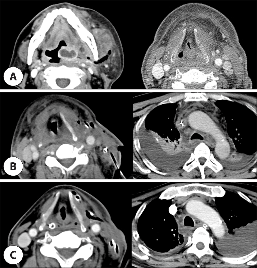
환자는 위중증 COVID-19 확진환자 진료지침에 따라 음압집중치료실에 입원하였다. COVID-19가 동반되어 있어 폐 기능이 저하될 가능성을 고려하여, 입원 당일 집중치료실에서 기관절개술 및 인공호흡기치료를 시작하였고 입원 2일째 음압수술실에서 절개배농을 시행하였다. 경부 제2, 3, 4, 5구역에 걸쳐 악취를 동반한 조직액 및 근막의 광범위한 괴사 소견이 관찰되어 경부의 괴사성 근막염으로 진단하고, 괴사 조직을 절제하고 여러 차례 세척한 뒤 3개의 배액관을 삽입하였다. 절개부는 봉합을 하지 않고 개방된 상태로 유지하며, 하루에 3회 이상 큐렛을 이용하여 괴사 조직을 제거하고 세척하는 한편, 광범위 정맥 항생제를 함께 투여하였다. 입원 5일째 혈액검사상 CRP 14.66, AST 66, ALT 54, BUN 38.6, Cr 0.77로 다발장기부전은 호전 추세를 보였으나(Table 1), 창상으로는 근막의 광범위한 괴사가 지속되고 있었으며 입원 8일째 시행한 전산화단층촬영 영상상에서도 경부에서부터 종격동까지 저음영 및 공기음영이 연장되어 있으면서 양측 흉수가 나타났다(Fig. 1B). 흉수천자를 통해 다량의 장액성 액체를 배액하였고, 흉수에 대한 검사상 백혈구 5,695, 세균배양검사상 특이 균이 배양되지 않아 다발장기부전과 관련된 삼출성 흉수로 생각되었다. 하행성 괴사성 종격동염으로의 진행에 대해 흉부외과 협진을 시행하였고, 환자의 전신상태를 고려하여 수술보다는 내과적 치료를 권유받아 감염내과와 지속적인 협진하에 경부의 개방창상을 통한 세척 및 괴사조직 절제와 항생제 치료를 지속하였다. 입원 16일째, 환자는 COVID-19 확진 후 21일이 경과하여 일반집중치료실로 전실하였고, 혈액검사 및 수술창 부위, 경부전산화단층촬영 영상 모두에서 점차 호전을 보여(Fig. 1C) 입원 21일째 배액관을 유지하고 절개부위를 봉합하였으며, 입원 33일째 일반 병실로 전실 후, 39일째 모든 배액관을 제거하였다. 환자는 장기간의 집중치료실 치료로 인한 전신위약에 대한 재활치료 후 입원 145일째 퇴원하였다.

고찰

COVID-19는 코로나바이러스의 한 종류인 SARS-CoV-2에 의해 발생하는 호흡기계의 바이러스성 감염병이며,3) 괴사성 근막염은 표재성 근건막계 및 천근막층을 침범하는 세균성 감염으로1) 두 질환의 발병기전은 차이가 있으나, COVID-19와 심경부감염이 동반된 증례가 국외에서 몇 차례 보고된 적이 있다(Table 2).49) 모든 증례에서 심경부감염은 COVID-19와 동시에 진단되거나 COVID-19 진단 후 수 주 이내에 진단되었다. 농양이 편도주위공간에만 국한되었던 한 증례를 제외하고, 모든 증례에서 1회 이상의 수술적 치료가 필요하였으며, 세 증례에서는 기도 확보를 위한 기관절개술이 시행되었다. 그러나 국내에서는 아직 관련된 보고는 없었다.

Table 2. Literature review of deep neck infection with COVID-19
Reference Sex/age Medical history Symptom Time sequence with COVID-19 diagnosis Treatment Prognosis
Orhan et al.4) M/64 Sore throat, left neck and chin pain Simultaneously Antivirals
Intravenous antibiotics
Steroid
Surgical drainage
Tracheostomy
Expired
Awobajo et al.5) F/60 Hypertension
Hyperlipidemia
Diabetes
Chronic kidney disease
Sore throat, right neck pain, voice change, odynophagia, drooling, dyspnea 3 weeks after Intravenous antibiotics
Surgical drainage (4 times)
Tracheostomy
Rehabilitation for dysphagia
Özer et al.6) M/45 Fever, general weakness, trismus, left chin and neck swelling After (being quarantined of COVID-19) Steroid
Intravenous antibiotics
Surgical drainage
Tracheostomy
Discharged
Shiraishi et al.7) M/55 Hypertension
Sleep apnea
Left shoulder pain After 12 days (being admissioned for treatment of COVID-19) Antivirals
Intravenous antibiotics
Steroid
Surgical drainage
Discharged
Sideris et al.8) M/21 Sore throat, fever, odynophagia, voice change Simutaneously Intravenous antibiotics
Steroid
Discharged
Charlton et al.9) F/- Simutaneously Intravenous antibiotics
Bedside drainage (2 times)
Discharged
Download Excel Table

중증 COVID-19에서 적용될 수 있는 인공호흡기치료 등에 의해 이차적인 상기도감염의 발생 가능성이 올라갈 수 있고,10) 당뇨와 같은 기저질환이 있는 환자의 경우 심경부감염에 대해 보다 취약하면서 COVID-19의 중증도도 올라갈 수 있으나,11,12) 아직까지 COVID-19와 괴사성 근막염의 인과관계는 불분명하다. 그러나 COVID-19의 증상이 괴사성 근막염과 같은 심경부감염의 원인이 되는 상기도 감염의 증상과 유사하기 때문에 병의 발견이 지연될 수 있고,13) COVID-19의 치료과정에서 사용되는 스테로이드와 면역억제제로 인해 심한 염증성 질환의 진단이 어려울 수 있다. 이와 더불어 COVID-19로 확진 시에 위중증으로 분류되지 않는다면 자가에서 격리하게 되는데,2) 이로 인해 의료기관으로의 접근성이 떨어질 수 있으며 이것은 병의 발견을 지연시켜 보다 많은 합병증을 유발하는 원인이 될 수 있다.9) 따라서, 괴사성근막염의 사망률이 높고 그 예후가 치료의 시작 시점과 밀접하게 연관되어 있다1)는 점을 고려하면 COVID-19는 괴사성 근막염의 진단을 지연시켜 예후를 불량하게 만드는 요인이 될 수 있다. Charlton 등에 따르면 COVID-19 대유행시기에 심경부감염으로 진단받은 환자들은 그렇지 않은 환자들에 비해 약 2.7일 늦게 의료기관을 방문하였으며, 질병의 중증도 및 합병증 발생률도 증가하여 기관절개술까지 필요한 경우도 7배 높은 것으로 나타났다.9)

한편 본 환자는 입원 당시에도 응급 수술의 필요성은 인정되었으나, 원내 음압 수술실의 개수가 제한되어 대기 시간이 길어졌고 입원 2일째 절개배농을 시도하였다. 환자는 하루 3회 이상 개방 창상을 통한 괴사조직 제거 및 세척을 시행하였고 이를 위해 하루 3번 이상, 2명 이상의 의료진이 개인 보호장구를 갖춘 뒤 집중치료실에 출입하여야 했다. 따라서 COVID-19가 동반되지 않은 경우에 비해 많은 인적 및 시간적 자원이 필요하였고, 상처 소독에 있어서 필요한 기구 등을 즉시 조달 받기에 어려움이 있었다. 2000년 이후 세계적인 바이러스 질환의 대유행은 약 5-6년 주기로 반복되고 있으며,3) 가까운 미래에 또다른 신종감염병의 범세계적인 유행이 발생할 가능성도 배제할 수 없다. 따라서 이번 COVID-19 대유행 시기에 여러 환자들을 치료한 경험을 토대로, 의료자원을 적절히 분배하고 신종감염병 프로토콜을 정립하여, 또 다른 신종감염병의 범유행시기에 대비하여야 한다. 본 환자처럼 격리가 필요한 경우, 음압상처치료 등을 적용함으로써 환자의 통증과 번거로움을 줄이고 상처 소독에 소요되는 시간도 크게 줄일 수 있으며1) 감염병환자와의 접촉 시간을 줄여 질병 전파의 위험성도 함께 낮출 수 있다. 또한 본 환자처럼 즉각적인 절개배농이 어려운 경우, 침상에서 기관절개술을 시행한 뒤 초음파 유도하 배농술을 먼저 시행하는 것도 대안이 될 수 있다.14) 기도 폐색의 위험성이 크지 않으면서 전신마취를 통한 절개배농이 어려운 심경부 감염 환자에서, 초음파 유도하 배농술로 효과적으로 치료한 경우가 여러 차례 보고된 바 있으며,14) 경과에 따라 추후 수술적 배농이 가능할 때 절개배농을 계획할 수 있다.

또한 환자들이 신종감염병 진단 후 격리가 되거나, 신종감염병에 대한 우려로 대외활동을 자제하게 되면서, 신종감염병의 범유행시기에 의료 체계로의 접근성이 떨어진다는 문제점도 보고되었다.9) 이에 대해 빠른 시일 내에 치료가 필요한 환자들이 치료 시기를 놓치지 않도록 신종감염병 대유행시기에 의료 체계 접근성을 보완하기 위한 대책이 함께 필요하다. 이번 COVID-19 대유행시기에 나타났던 이러한 문제점들을 개선하고 보완하여 명확한 대응 체계를 정립해 둔다면, 미래에 새로운 감염병이 유행하더라도 잘 대처할 수 있으리라 생각한다.

Acknowledgements

Not applicable.

Funding Information

Not applicable.

Conflicts of Interest

No potential conflict of interest relevant to this article was reported.

Author Contribution

Conceptualization: Lee G.

Data curation: Kwon E, Lee G.

Formal analysis: Lee G.

Methodology: Kwon E, Lee G.

Validation: Lee G.

Investigation: Kwon E.

Writing - original draft: Kwon E.

Writing - review & editing: Kwon E, Lee G.

Ethics Approval

Not applicable.

References

1.

Lee S, Kim H, Kwon TK. A case of negative pressure treatment on necrotizing fasciitis. Korean J Otorhinolaryngol Head Neck Surg 2012;55(12):791-4.

2.

Central Disease Control Headquarters, Central Disaster Management Headquarters. Guidelines in response to coronavirus disease 2019. 12nd ed. [Internet]. 2022 [cited 2023 Oct 10]. Available from: https://www.cdc.go.kr/board/board.es?mid=a20507020000&bid=0019&act=view&list_no=366558&tag=&nPage=12020

3.

Jin YJ, Kim SD. Tracheostomy in COVID-19 pandemic era. Korean J Otorhinolaryngol Head Neck Surg 2021;64(9):613-8.

4.

Orhan A, Karaketir S, Özdemir M, Akmeşe A. An unusual presentation of a patient with Covid-19 infection—Lemierre’s syndrome. Turk J Ear Nose Throat 2022;32(3):68-70.

5.

Awobajo MD, Agarwal AN, Hackman SD. Retropharyngeal abscess: an unusual complication in a COVID-19 patient. Otolaryngol Case Rep 2021;20: 100312.

6.

Özer F, Alkan Ö, Akin Ş. Sudden deep neck abscess formation in a patient with COVID-19. Turkiye Klinikleri J Case Rep 2021;29(3):120-4.

7.

Shiraishi K, Kasai H, Saito M, Kawaguchi H, Kinoshita T, Suzuki T, et al. Case of a deep neck abscess during treatment for COVID-19. Am J Case Rep 2022; 23:e936034.

8.

Sideris AW, Ghosh N, Lam ME, Mackay SG. Peritonsillar abscess and concomitant COVID-19 in a 21-year-old male. BMJ Case Rep 2020;13(9):e238104.

9.

Charlton AR, Simon R, Simons A. COVID-19 pandemic—impact on deep neck space infections: a retrospective cohort study. Clin Otolaryngol 2023;48(3): 479-88.

10.

Clancy CJ, Nguyen MH. Coronavirus disease 2019, superinfections, and antimicrobial development: what can we expect? Clin Infect Dis 2020;71(10):2736-43.

11.

Singh AK, Gupta R, Ghosh A, Misra A. Diabetes in COVID-19: prevalence, pathophysiology, prognosis and practical considerations. Diabetes Metab Syndr 2020;14(4):303-10.

12.

Vieira F, Allen SM, Stocks RM, Thompson JW. Deep neck infection. Otolaryngol Clin North Am 2008;41(3): 459-83.

13.

El-Anwar MW, Elzayat S, Fouad YA. ENT manifestation in COVID-19 patients. Auris Nasus Larynx 2020; 47(4):559-64.

14.

Han KY, Lee JH, Kim JH, Kim JY, Shim BS, Song YJ. Ultrasonography guided percutaneous aspiration and drainage of deep neck abscesses. Korean J Otorhinolaryngol Head Neck Surg 2009;52(4):354-8.