Journal of Clinical Otolaryngology Head and Neck Surgery
The Busan, Ulsan, Gyeoungnam Branch of Korean Society of Otolaryngology-Head and Neck Surgery
Case Report

청소년 환자에서 발생한 비인두 성숙 기형종 1례

박혜진1https://orcid.org/0000-0003-4237-6834, 박지환1https://orcid.org/0000-0001-9301-6957, 최경운2https://orcid.org/0000-0002-3848-1781, 조규섭1,*https://orcid.org/0000-0002-4381-6996
Hye-Jin Park1https://orcid.org/0000-0003-4237-6834, Ji-Hwan Park1https://orcid.org/0000-0001-9301-6957, Kyung-Un Choi2https://orcid.org/0000-0002-3848-1781, Kyu-Sup Cho1,*https://orcid.org/0000-0002-4381-6996
1부산대학교 의과대학 이비인후과학교실
2부산대학교 의과대학 병리학교실
1Department of Otorhinolaryngology and Biomedical Research Institute, Pusan National University School of Medicine, Pusan National University Hospital, Busan, Korea
2Department of Pathology and Biomedical Research Institute, Pusan National University School of Medicine, Pusan National University Hospital, Busan, Korea
*Corresponding author: Kyu-Sup Cho, Department of Otorhinolaryngology and Biomedical Research Institute, Pusan National University School of Medicine, Pusan National University Hospital, Busan 49241, Korea, Tel: +82-51-240-7824, Fax: +82-51-246-8668, E-mail: choks@pusan.ac.kr

© Copyright 2023 The Busan, Ulsan, Gyeoungnam Branch of Korean Society of Otolaryngology-Head and Neck Surgery. This is an Open-Access article distributed under the terms of the Creative Commons Attribution Non-Commercial License (http://creativecommons.org/licenses/by-nc/4.0/) which permits unrestricted non-commercial use, distribution, and reproduction in any medium, provided the original work is properly cited.

Received: Dec 11, 2023; Revised: Dec 20, 2023; Accepted: Dec 22, 2023

Published Online: Dec 31, 2023

ABSTRACT

Teratomas are congenital tumors originating from one or more germ layers and are very rare in the head and neck. They may be histologically mature and oncologically benign. Specifically, teratomas derived from the nasopharynx are even rarer and are typically discovered after childbirth or in early childhood, causing respiratory obstruction and difficulty in breastfeeding. We report and review a case of nasopharyngeal mature teratoma in an adolescent patient. This suggests that when a nasopharyngeal mass is found in adolescents and young adults, the possibility of a teratoma should be considered.

Keywords: Nasopharynx; Teratoma; Adolescent

서론

기형종은 외배엽, 중배엽 및 내배엽으로 구성된 세 가지 생식 세포층 가운데 하나 이상의 배엽에서 기원한 선천성 종양이다. 조직학적으로 피부, 모발, 뼈, 연골 등으로 구성되면 성숙 기형종으로, 신경 상피 조직과 같은 미성숙 부분의 구성 비율이 높으면 미성숙 기형종으로 분류된다.1) 주로 몸의 정중선을 따라 생식선, 천미골(sacrococcyx), 종격동에 호발하지만 드물게 두경부 영역에 발생하기도 한다.2) 성인보다 소아에서 발생 빈도가 더 높으며, 소아 기형종의 유병률은 20,000–40,000명 중에 1명으로 비교적 드물다. 전체 기형종 가운데 두경부 영역에서의 유병률은 2%–5%인데 경부 기형종이 가장 흔하며, 비인두 기형종이 그 다음 순이다.3,4) 일반적으로 비인두 기형종은 신생아기에 호흡 곤란 및 수유 불가와 같은 상기도 폐쇄 증상을 일으켜 발견되는 경우가 흔하나, 크기와 발생 위치에 따라 증상은 달라질 수 있다.5)

신생아나 성인에서 발견된 비인두 기형종의 경우 국내에 보고된 바가 있지만,6,7) 청소년기에 발견된 비인두 성숙 기형종은 국내에 보고된 바가 없다. 저자들은 우연히 발견된 비인두 종물로 수술 받은 17세 남자 환자가 비인두 성숙 기형종으로 진단받은 1례를 경험하였기에 문헌고찰과 함께 보고하는 바이다.

증례

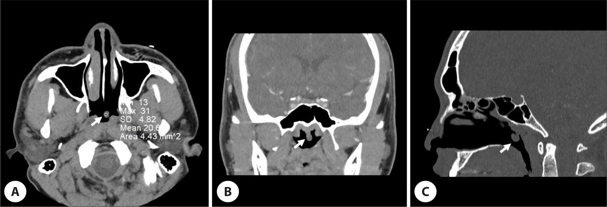
평소 기저질환이 없는 17세 남아가 1달 전 독감에 걸려 개인병원에서 치료받던 중, 비인강경 검사상 비인두 종물이 발견되어 수술적 치료를 권유 받고 본원 이비인후과 외래로 내원하였다. 비인강경 검사상 비인두에 경계가 명확한 무통성의 유경형 종물이 관찰되었다(Fig. 1A). 수술 전 시행한 조영 증강 부비동 전산화 단층촬영(paranasal sinus computed tomography, PNS CT)에서 비인두 중앙에 인두강쪽으로 자란 약 8×6 mm 크기의 조영 증강되지 않는 유경형 종괴가 확인되었다. 종괴는 뇌실질 및 뇌척수액과의 교통이 없고 경계가 분명하였으며, 조영 증강 전 CT에서 HU(hounsfield unit)이 평균 20.6으로 단백 성분이 포함된 양성 낭성 종물일 것으로 추정되었다(Fig. 2).

jcohns-34-4-231-g1
Fig. 1. Preoperative and postoperative endoscopic findings. A: Preoperative endoscopic image shows a well-defined protruding mass in the nasopharynx. B: Postoperative endoscopic image shows that the nasopharyngeal mass has been completely removed and the area is healing well.
Download Original Figure
jcohns-34-4-231-g2
Fig. 2. Preoperative paranasal sinus computed tomography (CT). Contrast-enhanced CT images show a round well defined non-enhancing mass (white arrow) in the nasopharynx on axial (A), coronal (B), and sagittal view (C).
Download Original Figure

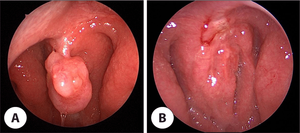
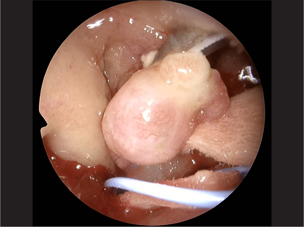
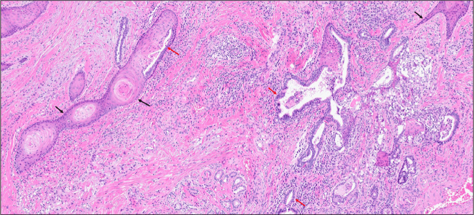
전신마취 하에 내시경 비인두 종물 제거술을 시행하였다. 비인두의 중앙에 위치한 유경형 종물로 안전역을 확보하여 종양을 일괄 절제하기 위해 Harmonic scalpel(Ethicon Endo-Surgery, Cincinnati, OH, USA)로 종괴의 기시부를 비인두벽에 최대한 가까이 붙여 절제하고, 기시부를 소작한 후 수술을 종료하였다(Fig. 3). 출혈은 거의 없었으며, 합병증 없이 수술 후 다음날 퇴원하였다. 조직검사에서 현미경 소견상 단순 및 중층 원주 상피 세포와 편평 상피 세포로 둘러싸인 다방성의 낭성 구조물로, 비인두에 발생한 성숙 기형종으로 진단되었다(Fig. 4). 수술 2주 후 국소 소견상 특이 소견이 관찰되지 않았으며 현재까지 재발없이 경과 관찰 중이다(Fig. 1B).

jcohns-34-4-231-g3
Fig. 3. Intraoperative endoscopic findings. The nasopharyngeal mass was approximately 8×6 mm in size and was totally resected using harmonic scalpel via an endoscopic approach.
Download Original Figure
jcohns-34-4-231-g4
Fig. 4. Histopathological finding of nasopharyngeal mass. Microscopic examination showed numerous spaces lined by simple and stratified columnar (red arrow) and squamous epithelia (black arrow), which was suggestive of mature teratoma (hematoxylin and eosin, ×100).
Download Original Figure

고찰

기형종은 한 종류 이상의 배엽에서 기원한 선천성 종양으로 구성 배엽의 수나 분화도에 따라 진성 기형종, 유피종, 유기형종 등으로 분류되며, 진성 기형종은 분화도와 미성숙 조직의 비율에 따라 성숙 기형종, 미성숙 기형종, 악성화 된 기형종으로 구분할 수 있다.1,8,9) 기형종은 신생아 및 영아기에 90%이상 발견되고 조직학적으로는 양성이 대부분이다. 하지만 소아 기형종 가운데 2.5%에서 악성으로 진단되며 성인에서 발견된 경우 69%가 악성으로 진단되므로, 나이가 증가할수록 기형종의 악성화 가능성이 높아진다고 볼 수 있다. 악성 기형종의 위험도는 조직의 미분화 정도보다는 나이와의 관련성이 더 높은 것으로 알려져 있으므로,10) 청소년기 이후의 성인에서 진단될 경우 악성화 가능성을 항상 염두해 두어야 한다. 본 증례는 17세의 청소년기에 발견된 비인두 성숙 기형종으로 일반적으로 기형종이 잘 발생하는 연령대보다 높은 연령층에서 발생하였으나 양성으로 진단되었다.

두경부 영역의 기형종은 전체 기형종 가운데 약 2%–5%를 차지하고, 유병률은 경부, 비인두 순이다.3,4) 비인두 기형종은 비인두의 후벽이나 측벽에 발생하는 경우가 많다. 비인두는 발생과정에서 외배엽과 내배엽 점막층 사이의 접합부에서 형성되기 때문에, 기형종의 발생 가능성이 상대적으로 높다.11) 비인두의 기형종 진단에는 CT, 자기 공명 영상 장치(magnetic resonance imaging) 등이 유용하며 고형 및 낭성 성분의 분포 비율, 지방이나 석회화 등을 확인함으로써 진단에 도움을 얻을 수 있다. 비인두에 발생하는 기형종과 감별해야 할 질환으로는 뇌류(encephalocele), 척삭종(chordoma), 하마종(hamartoma), 신경아교종(glioma), 횡문근종(rhabdomyoma), 림프관종(lymphagioma), 두개인두종(craniopharyngioma) 등이 있다. CT로 감별할 수 있는데, 뇌류의 경우 두개저의 미란이 관찰되는 반면, 기형종은 두개 내강과의 연결이 없고 골미란의 소견도 관찰되지 않는다.12,13) 본 증례에서 수술 전 시행한 PNS CT상 비인두에서 발생한 조영 증강되지 않는 매끈한 경계의 낭성 유경형 종괴가 뇌실질 및 뇌척수액과의 교통이 없어 뇌류와 감별되었다. 단백 성분이 포함된 양성의 낭성 종물일 것으로 추정되었으나, 치아와 같이 기형종을 강력히 시사하는 석회화 된 조직은 확인되지 않아 영상 소견으로 기형종을 의심하기는 어려웠다.

성인의 경우와는 달리 소아의 두경부에 생긴 기형종은 조직 병리학적으로 양성인 경우가 대부분이지만, 종괴의 위치와 크기에 따라 상기도 폐쇄를 초래할 수 있다. 이로 인해 호흡 곤란, 수유 장애 및 코골이 등의 증상이 나타날 수 있으며, 종괴가 긴 경우에는 후두를 막거나 자극하여 기침, 구토, 청색증을 유발하기도 한다.14,15) 본 증례의 환자는 증상 없이 우연히 비인두 종물이 발견된 경우로, 종괴의 크기가 작고, 인두강 쪽으로 종괴의 일부분만이 돌출되어 상기도 폐쇄나 자극을 일으키지 않았기 때문으로 생각할 수 있다.

본 증례는 조직 병리학적으로 양성 성숙 기형종으로 진단되었다. 일반적으로 성숙 기형종은 양성이며, 완전히 분화된 조직인 털, 땀샘, 지방조직, 치아, 손톱, 신경, 연골 및 근육 등을 포함하고 있는 경우가 많다. 또한 주변 구조물을 침범하거나 전이되지 않는 것으로 알려져 있어, 수술적 완전 절제가 치료 원칙이다.1) 완전 절제 후 재발은 드물며, 예후는 좋은 편이다. 하지만 불완전하게 절제된 경우 드물지만 재발할 수 있으며,14) 성숙 기형종이라 할지라도 연령 증가에 따라 악성화 가능성이 있기 때문에 조기에 진단하여 완전 절제하는 것이 중요하다고 할 수 있다.10) 본 증례에서 유경형 종물의 기시부를 Harmonic scalpel을 이용하여 최대한 남김없이 절제하였으며, 비인두의 기시부와 주변부를 소작하여 불완전 절제의 가능성을 최소화 하였다. 하지만 향후 드물지만 재발할 여지가 있기 때문에 추적 관찰이 지속되어야 할 것으로 생각된다. 악성 기형종에서 종양이 난황난종양(yolk sac tumor)이나 융모상피암(choriocarcinoma)의 조직을 가지고 있다면, 혈액검사에서 알파 태아단백질(alpha-fetoprotein, αFP)이나 베타-인간 융모성 성선자극호르몬(beta-human chorionic gonadotrophin)의 혈중 수치가 증가할 수 있다. αFP의 경우 기형종의 악성 전환의 지표로 보기에는 민감도가 낮아 초기 진단에는 유용성이 떨어질 수 있지만,16) 재발 여부의 판단에 도움이 될 수 있다. 따라서, 향후 재발의 평가에 비인강경 및 영상 검사 이외에도 혈액 검사를 시행해 볼 수 있을 것이다.

본 증례는 청장년층에서 비인두 종물이 발견된 경우, 드물지만 기형종의 가능성을 고려할 수 있으며 진단과 함께 완전한 수술적 절제가 조기에 시행되는 것이 중요하다는 것을 시사한다고 하겠다.

Acknowledgements

Not applicable.

Funding Information

Not applicable.

Conflicts of Interest

No potential conflict of interest relevant to this article was reported.

Author Contribution

Conceptualization: Cho KS.

Data curation: Park JH.

Methodology: Choi KU.

Writing - original draft: Park HJ.

Writing - review & editing: Park HJ, Park JH, Choi KU, Cho KS.

Ethics Approval

This manuscript was approved by the Institutional Review Board of the Pusan National University Hospital (Approval No. 2312-012-134).

References

1.

Carr MM, Thorner P, Phillips JH. Congenital teratomas of the head and neck. J Otolaryngol 1997;26(4):246-52.

2.

Byard RW, Jimenez CL, Carpenter BF, Smith CR. Congenital teratomas of the neck and nasopharynx: a clinical and pathological study of 18 cases. J Paediatr Child Health 1990;26(1):12-6.

3.

Holt GR, Holt JE, Weaver RG. Dermoids and teratomas of the head and neck. Ear Nose Throat J 1979; 58(12):520-31.

4.

Bergé SJ, von Lindern JJ, Appel T, Braumann B, Niederhagen B. Diagnosis and management of cervical teratomas. Br J Oral Maxillofac Surg 2004;42(1):41-5.

5.

Zia-ul-Miraj Ahmad M, Brereton RJ, Madden NP. Oronasopharyngeal teratomas. Pediatr Surg Int 1996;11(2-3):203-5.

6.

Yoo DK, Song CJ, Ahn YS, Kang DY. Nasopharyngeal teratoma in an adult: a case report. J Korean Radiol Soc 2002;47(2):161-4.

7.

Eom JW, Hwang MS, Kang MS. A case of concurrent mature teratoma and heterotopic gastrointestinal cyst in head and neck. Korean J Otolaryngol 2003;46(6):536-9.

8.

El-Sayed Y. Teratoma of the head and neck. J Laryngol Otol 1992;106(9):836-8.

9.

Batsakis JG. Teratomas of the head and neck. In: Batsakis JG. Tumors of the head and neck. 2nd ed. Baltimore: Williams & Wilkins; 1979. p.226-32.

10.

Jordan RB, Gauderer MWL. Cervical teratomas: an analysis. Literature review and proposed classification. J Pediatr Surg 1988;23(6):583-91.

11.

Zarbo RJ, McClatchey KD. Nasopharyngeal hamartoma: report of a case and review of the literature. Laryngoscope 1983;93(4):494-7.

12.

Handler SD, Beverly Raney R Jr. Management of neoplasms of the head and neck in children. I. Benign tumors. Head Neck Surg 1981;3(5):395-405.

13.

Snow JB Jr. Neoplasms of the nasopharynx in children. Otolaryngol Clin North Am 1977;10(1):11-24.

14.

Coppit III GL, Perkins JA, Manning SC. Nasopharyngeal teratomas and dermoids: a review of the literature and case series. Int J Pediatr Otorhinolaryngol 2000;52(3):219-27.

15.

Kountakis SE, Minotti AM, Maillard A, Stiernberg CM. Teratomas of the head and neck. Am J Otolaryngol 1994;15(4):292-6.

16.

Touran T, Applebaum H, Frost DB, Richardson R, Taber P, Rowland J. Congenital metastatic cervical teratoma: diagnostic and management considerations. J Pediatr Surg 1989;24(1):21-3.