Introduction
Hemangiomas of the head and neck are known to be common, but they are found to be rare in the sinonasal cavity. Most of the sinonasal hemangiomas are capillary type hemangiomas and originate from the nasal septum or vestibule.1)
The authors report a case of a mass in the left nasolabial fold that was diagnosed as hemangioma after resection using trans-nasal approach along with a review of the literature.
Case
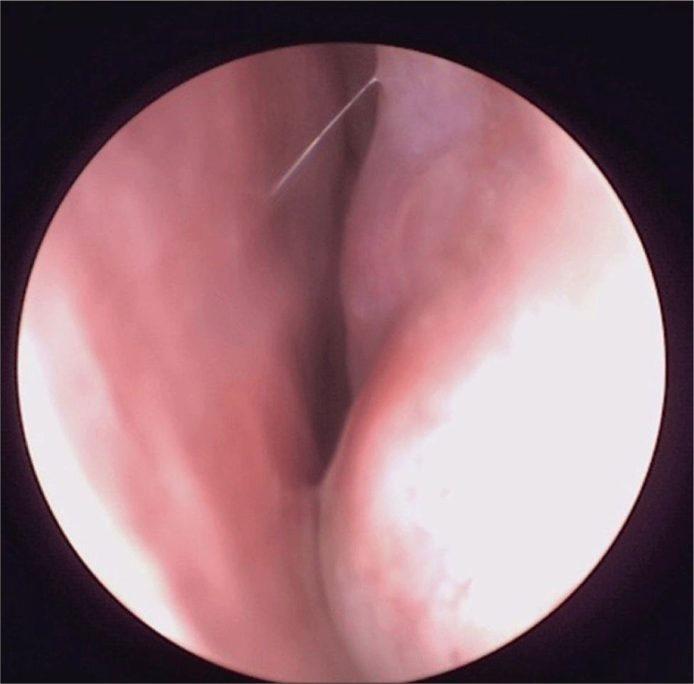
A 54-year-old female patient presented with a complaint of left sided nasal obstruction. The patient had no significant medical history other than the use of hormone medication. She had been experiencing nasal obstruction for a while, but experienced worsening one week ago. She denied any other symptoms such as rhinorrhea or epistaxis. Physical examination showed a soft mass about a size of 1.5×1.5 cm at the nasolabial fold area, which was palpable but no signs of tenderness. Endoscopic findings showed septal deviation to left and edematous changes at the nasolabial area (Fig. 1).

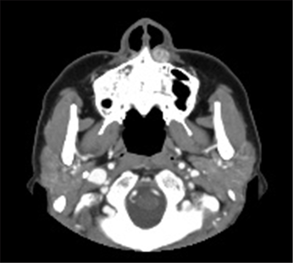
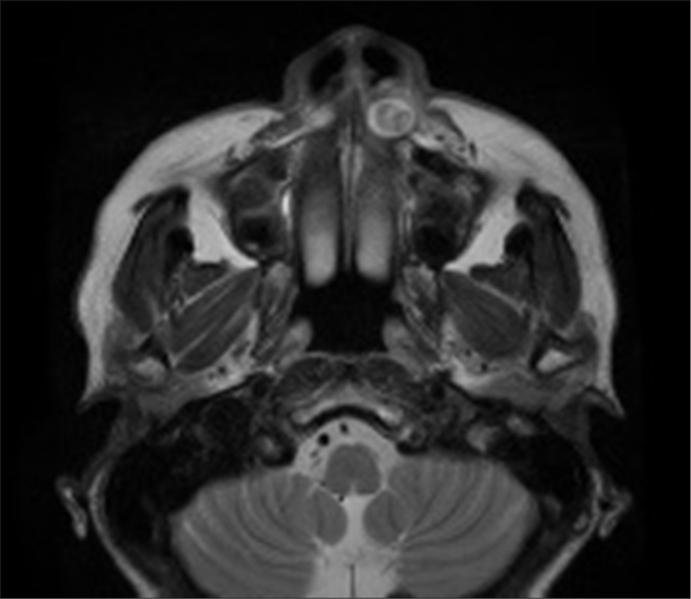
To determine the location and extent of the lesion, contrast-enhanced paranasal sinus computed tomography was done. 1.3×0.9×1.3 cm sized heterogeneously enhancing mass was found at the nasolabial area. Surrounding bone erosion was observed and no calcifications were found within the lesion (Fig. 2). Considering the patient’s medical history and examination findings, possible benign soft tissue origin tumors such as nasolabial cyst or hemangioma were considered. Biopsy was performed during an outpatient visit which showed chronic inflammation. Prior to surgery, paranasal sinus magnetic resonance imaging (MRI) was done for additional evaluation of the mass. 1.3×0.9×1.3 cm sized, peripheral rim enhancing mass, surrounding bone remodeling was observed and no diffusion weighted image (DWI) restriction was seen. T2 weighted image showed a round mass with heterogenic signal intensity within the mass (Fig. 3). Based on these findings, nasolabial cyst and hemangioma were considered and a plan was made for left nasal mass removal by intranasal or sublabial approach.


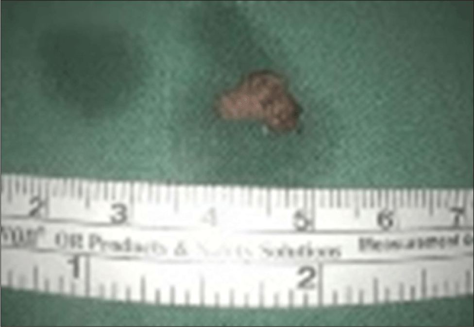
After receiving the prescribed antibiotics after the biopsy performed on January 3rd, patient developed diarrhea and was admitted to the Department of Infectious Diseases and started oral vancomycin treatment. During hospitalization, diarrhea symptoms improved, so the patient transferred to the Otorhinolaryngology department on January 16th for surgery scheduled on the following day. The surgery was performed on general anesthesia using a trans-nasal approach. After making an incision at the left nasal floor mucosa, flap was raised and the mass was dissected. Mass was found to be adhered to the maxillary bone anterior nasal spine. After the removal of the main mass (Fig. 4), the remaining mass was completely excised using a debrider. Histopathological examination showed a mass mainly composed of vessels leading to the final diagnosis of hemangioma (Fig. 5). According to the Pathology department, the hemangioma subtype was not clearly able to be confirmed, however, the findings are consistent with characteristics resembling a venous hemangioma. The patient is currently under postoperative follow-up observation.

Discussion
Hemangioma is the most common congenital benign tumor, over 50% cases occurring in the head and neck region.2,3) It is known to primarily occur congenitally, but can also be caused by errors in vascular formation, hemodynamic abnormality, and local trauma.4,5) Approximately 95% of cases develop within the first 6 months of birth, and undergo natural regression by the age of 12.6) However, hemangiomas persisting beyond that age are known to have difficulty undergoing natural regression.6) According to the WHO classification of hemangiomas, they can be classified as cavernous, capillary, arteriovenous, venous, intramuscular, synovial hemangiomas based on their histopathology.7) Cavernous hemangiomas consist of dilated large vessels or sinusoidal blood space. Due to its low rate of regression, incidence rate of cavernous hemangioma is higher in adults compared to capillary hemangioma. Capillary hemangiomas consist of capillary sized vessels with flattened endothelial cells. Mostly they are found at childhood and spontaneous regression is common.8) Venous hemangiomas consist of veins with small thick walls and elastic plate and fiber rarely exists inside. This feature makes it possible to distinguish venous hemangiomas with other types of hemangiomas.4) In our case, histopathologic characteristics of the hemangioma did not follow cavernous nor capillary. According to the Pathology department, a definitive conclusion is difficult to be made but venous type can be considered. The authors concluded as venous hemangioma due to the features of thick walled vessels.
Diagnosis of hemangioma is carried out by careful history taking, medical and physical examination, and imaging studies such as computed tomography (CT), MRI, angiography.2,9) Symptoms of sinonasal hemangiomas include nasal obstruction, recurrent epistaxis, cheek swelling and bulging of the eyeball.10) Our patient presented with only nasal obstruction. CT shows a highly vascularized tumor, and shape, size relations with the surrounding structures, and extension to soft tissue.9,10) MRI shows a high signal intensity due to abundant blood flow on T2-weighted images. In the case with our patient, CT showed a heterogenous mass with bone remodeling and MRI T2-weighted image showed peripheral rim enhancement which was not the typical characteristics of the features above.
Differential diagnosis of hemangiomas in the sinonasal cavity include inverted papillomas, neuromas, polypoid masses mucoceles, or other benign or malignant vascular tumors.10) In our case, because of the location of the mass, nasolabial cyst was needed to be differentiated.
Treatment of hemangioma must consider patient age, the location and size, histopathologic characteristics of the hemangioma.3,6) Hemangiomas of the childhood mostly spontaneously involute, therefore can be observed.2,3,6) However in adults, spontaneous involution is rare, and surgical treatment must be considered. Prior to surgery, CT or angiographic evaluation must be performed due to massive bleeding risk during surgery.8,11) In order to prevent relapse, total resection of the mass is important.8) Nonsurgical treatments include local steroid injections which helps slowing the progression of the hemangioma. Pulsed dye laser, photocoagulation with laser also can be an effective method to reduce the volume of hemangioma.3)
In our case, due to the location of the mass, nasolabial cyst was the first impression to be considered. Also heterogenic enhancing characteristics found on computed tomography images, and T2-weighted images of MRI did not follow the standard characteristics of a hemangioma. Because of the bleeding risk during surgery, when encountering a nasolabial fold mass, hemangioma should be an impression to be considered. Previous studies report cases of intranasal hemangiomas, but cases of nasolabial fold hemangiomas were rare.12) Furthermore, the features of our case did not follow the typical characteristics of hemangiomas on imaging studies. Therefore, we report a rare case diagnosed as hemangioma in the nasolabial fold we have experienced.

