


Introduction
In 1936, Andre Lemierre reported throat infections, especially anaerobic septicemia after a peritonsillar abscess, and abscess formation in various areas, such as the lungs and joints.1) These findings are referred to as the Lemierre syndrome, which is characterized by thrombophlebitis of the internal jugular vein (IJV), septicemia, and septic emboli that spread to various organs, such as the lungs.1) Infectious bacteria are mainly anaerobic, and Fusobacterium necrophorum is the most common infectious bacterium causing Lemierre syndrome.
Despite the low incidence, Lemierre syndrome is a severe disease that progresses quickly, invades various organs, and causes septicemia. In the 2000s, the mortality rate is reported about 5%, which is significantly lower than the mortality rate of 90% initially reported in 1936.1,2) This drop in mortality is attributed to the development of antibiotics. Although the prevalence of Lemierre syndrome has been continuously declining since the advent of antibiotics, a recent spike has been observed. This increase is likely related to the increase in antibiotic-resistant bacteria.3) Therefore, understanding Lemierre syndrome is essential for the successful treatment of this disease despite the low number of cases encountered in clinical practice.
In this paper, we report the successful surgical drainage and postoperative treatment of a patient with Lemierre syndrome due to Streptococcus constellatus.
Case Report
A 51-year-old female patient complained of fever and left neck pain lasting for 1 week. She was admitted to another hospital and prescribed antibiotic treatment, but her symptoms did not improve and progressed to thrombocytopenia and septicemia. Therefore, she was transferred to the emergency room for further evaluation and treatment.
Swelling on the left side of the neck was observed on physical examination. Left peritonsillar swelling was observed on laryngoscopy, and her airway was patent. Her vital signs were as follows: body temperature, 37.7℃; systolic blood pressure, 70 mmHg; and diastolic blood pressure, 40 mmHg. Blood tests revealed thrombocytopenia (platelet count of 12×103/µL [reference: 140×103–400×103/µL]), leukocytosis (white blood cell count of 25.44×10³/µL [reference: 4.0×10³–11.0×10³/µL] and neutrophil segment of 92% [reference: 40%–73%]), and a high sensitivity C-reactive protein level of 20.31 mg/dL (reference: 0–0.5 mg/dL). The blood culture was positive for Clostridium clostridioforme approximately 2 days after admission. Therefore, septicemia due to an uncontrolled pharyngolaryngeal infection was suspected.
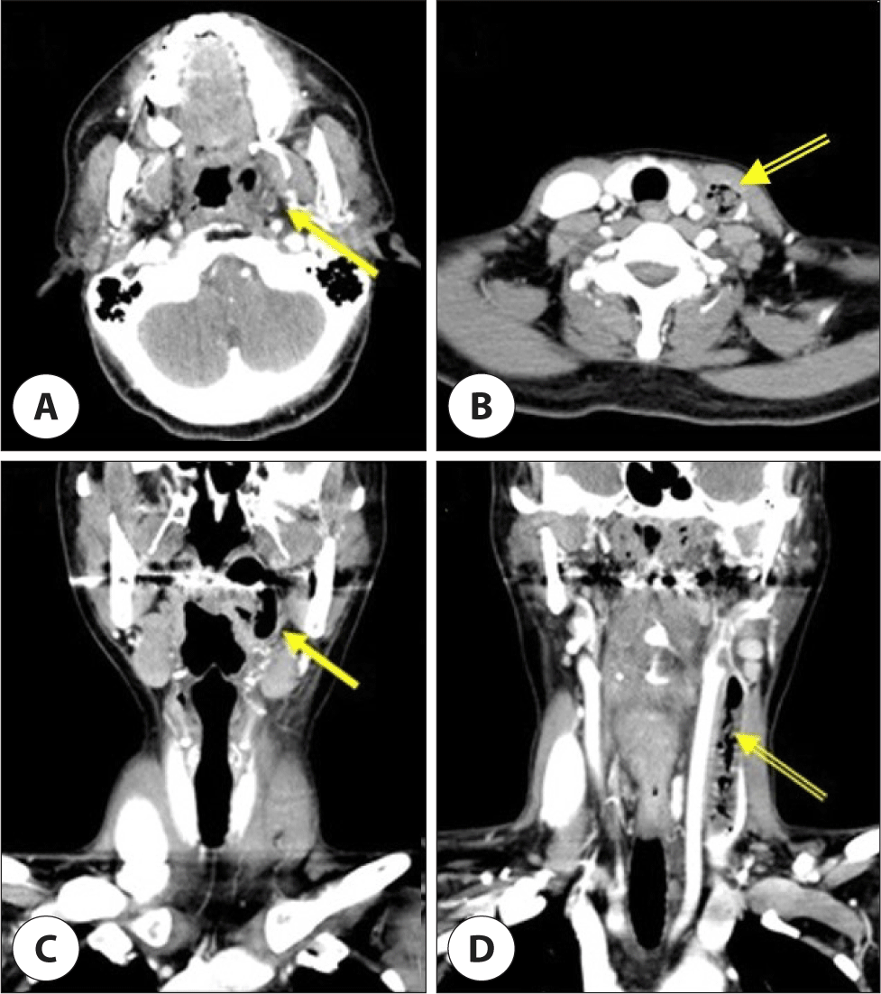
Contrast-enhanced computed tomography (CT) showed a peritonsillar abscess and thrombophlebitis of the left IJV (Fig. 1). As there was uncontrolled septicemia and thrombophlebitis of the IJV, we decided to perform a transcervical drainage rather than an intraoral drainage to obtain a wider surgical field and to remove the thrombosis inside IJV. Emergency abscess drainage was performed under general anesthesia. The abscess was observed along the left IJV from the mandible to the clavicle. The tearing of left facial vein was occurred during thrombectomy and facial vein was sutured with a 6-0 prolene.
Strepcococcus constellatus was cultured and it was susceptible to all antibiotics. Piperacillin/tazobactam and levofloxacin were used as antibiotics for 22 days after surgery.
After the surgery, the patient was admitted to the intensive care unit for treatment. A postoperative day 3 contrast-enhanced chest CT revealed diffuse airspace consolidations in the bilateral lower lobes and multiple patchy areas of glassy opacities of the lungs suggestive of septic pneumonia and bilateral pleural effusions. The pneumonia improved with continuous piperacillin/tazobactam and levofloxacin therapy. On postoperative day 8, she was transferred to a general ward and safely discharged on day 25.
Discussion
The onset of Lemierre syndrome is marked by oropharyngeal infection with symptoms such as a sore throat, fever, and cervical tenderness and swelling. Parapharyngeal abscess formation and peripheral suppurative thrombophlebitis in the IJV occur, causing complications such as septicemia, carotid artery rupture, and pulmonary embolism; these conditions affect other organs, such as the joints, kidneys and lungs.4)
The prevalence of Lemierre syndrome has gradually decreased due to the development of antibiotics, with an incidence between 0.6 and 2.3 per million people.4) In addition, the overall mortality rate is reported to be 4%–18%, 70% of patients are young adults between the ages of 16 and 25 years, and twice as many cases involve males compared with females.2,4) According to Karkos et al., approximately 200 papers on Lemierre syndrome were published from 1980 to 2010, and the number tended to increase with time, most likely due to increase in the number of antibiotic-resistant bacteria and newly developed diagnostic technology.3,5)
In general, Fusobacterium necrophorum is the most common bacteria causing Lemierre syndrome, accounting for about 62.0%–81.7% of cases. Streptococcus spp. and Staphylococcus spp. also cause Lemirre syndrome.4,6) In the present case, Streptococcus constellatus was cultured. Streptococcus constellatus was reported to cause neck abscesses and IJV thrombosis in 25% of Lemierre cases.7) It is reported that a mixed infection of Fusobacterium and Streptococcus is more dangerous than either alone.8) Therefore, if Streptococcus is identified, it is necessary to confirm that it is not co-infected with Fusobacterium. In this case, Streptococcus was identified as an isolated infection.
Diagnostic criteria for Lemierre syndrome have not yet been clearly established, but in 1989, Sinave et al. proposed diagnostic criteria based on characteristic symptoms: primary infection of the oropharynx, sepsis caused by one or more bacteria streaked on blood culture, IJV thrombosis, and involvement of other organ.9)
Contrast-enhanced CT is the most important tool for diagnosis, where abscess formation in the parapharyngeal area, a filling defect in the IJV, and septic pulmonary emboli can be observed. In addition to the IJV, there are cases where adjacent blood vessels, such as the superior ophthalmic vein, facial vein, and sigmoid sinus, are affected.10) The extent of the disease and the invading blood vessels need to be evaluated before surgical treatment.
Treatment of Lemierre syndrome is based on the use of antibiotics, and surgical treatment is performed when the lesion is localized and the patient's condition is operable. In general, penicillin, metronidazole, and second- and third-generation cephalosporins are used for 3–6 weeks or longer.11) However, as the number of antibiotic-resistant bacteria are increasing and bacteria other than Fusobacterium necrophorum can be an infection source, it is important to identify the strain of bacteria and determine the appropriate antibiotics.
As the main symptoms are of septic thrombophlebitis and septic emboli of the IJV, anticoagulants are sometimes used for treatment.12) Although many studies have been conducted on the use of anticoagulants, it is still unclear whether they are effective. After starting treatment, patients require an average hospitalization of about one month, and 58% of patients receive treatment in the intensive care unit.2)
In the case presented herein, the patient underwent immediate surgery with broad spectrum antibiotic treatment. Compared to 77.6% of immediate surgical drainage in patients with common deep neck infections, the rate of surgical drainage is relatively low for patients with Lemierre syndrome.8,13) Invasion and distant infection of the IJV and adjacent blood vessels and septicemia may limit the use of general anesthesia. Surgery helps with rapid recovery, but it is important to accurately grasp the extent of the lesion before the procedure.
Conclusion
Lemierre syndrome is rare but it could be fatal which requires prompt diagnosis and treatment. Contrast-enhanced CT is important to decide the extent of the lesion and culture-based antibiotics should be used immediately.
It seems that Streptococcus constellatus mono-infection is related to better prognosis combined with early surgical intervention including drainage and thrombectomy of IJV.