Case Report
익상구개와를 점유한 무증상의 세포성 신경초종의 우연한 진단과 제거 1예
An Incidental Diagnosis and Removal of an Asymptomatic Cellular Schwannoma Involving the Pterygopalatine Fossa: A Case Report
Hyeok Ro Kwon1
,
So Young Ko1,
Min Ah Kim2,
Jin Soon Chang1,*
Author Information & Copyright ▼
1Department of Otorhinolaryngology-Head and Neck Surgery, Veterans Health Service Medical Center, Seoul, Korea
2Pathology, Veterans Health Service Medical Center, Seoul, Korea
*Corresponding author: Jin Soon Chang, Department of Otolaryngology-Head and Neck Surgery, Veterans Health Service Medical Center, Seoul 05368, Korea, Tel: +82-2-2225-1384, Fax: +82-2-2225-1385, E-mail:
jsc4g@naver.com
© Copyright 2023 The Busan, Ulsan, Gyeoungnam Branch of Korean Society of Otolaryngology-Head and Neck Surgery. This is an Open-Access article distributed under the terms of the
Creative Commons Attribution Non-Commercial License (http://creativecommons.org/licenses/by-nc/4.0/) which permits
unrestricted non-commercial use, distribution, and reproduction in any
medium, provided the original work is properly cited.
Received: Oct 11, 2023; Revised: Nov 17, 2023; Accepted: Dec 11, 2023
Published Online: Dec 31, 2023
ABSTRACT
Schwannoma is a type of benign neurogenic tumor which may derive from the nerve sheath. They can occur throughout anybody but rarely manifest in the nasal cavity, representing less than 4% of all head and neck schwannomas. A 68-year-old woman presented to our clinic with a complaint of a mass occupying the space of the maxillary sinus beyond the pterygopalatine fossa. This mass was discovered during a brain magnetic resonance imaging (MRI) test conducted at another hospital during a routine health checkup. Paranasal sinus computed tomography (CT) and magnetic resonance imaging (MRI) revealed a heterogenous enhancing mass lesion measuring approximately 4.8×2.9×3.6 cm in size. We performed a mass excision via an endoscopic transnasal approach, and she was pathologically diagnosed with a cellular schwannoma. We present a didactic case report to highlight the minimal invasive technique and the absence of postoperative complications and recurrence, along with a relevant literature review.
Keywords: Schwannoma; Neurilemmoma; Pterygopalatine fossa; Maxillary sinus
서론
신경초종은 구형의 피막으로 둘러싸인 신경초세포에서 발생하는 양성 종양이다.1) 신경초세포가 없는 시신경과 후각 신경을 제외한 신경섬유의 신경초세포 어디서나 발생할 수 있지만, 전체 신경초종 중 비강 및 부비동에서 발생하는 빈도는 3%–4% 이하로 매우 드물다.2,3)
본 증례는 내원 1개월 정기 건강 진단에서 상악동까지 확장된 익상구개와와 공간의 종괴가 증상없이 우연히 뇌 자기공명영상 검사로 발견되어 본원으로 의뢰된 환자로 내시경하 비강 접근법을 통해 종물을 제거하고, 최종 조직검사상 세포성 신경초종으로 진단된 68세 여성을 문헌 고찰과 함께 보고하는 바이다.
증례
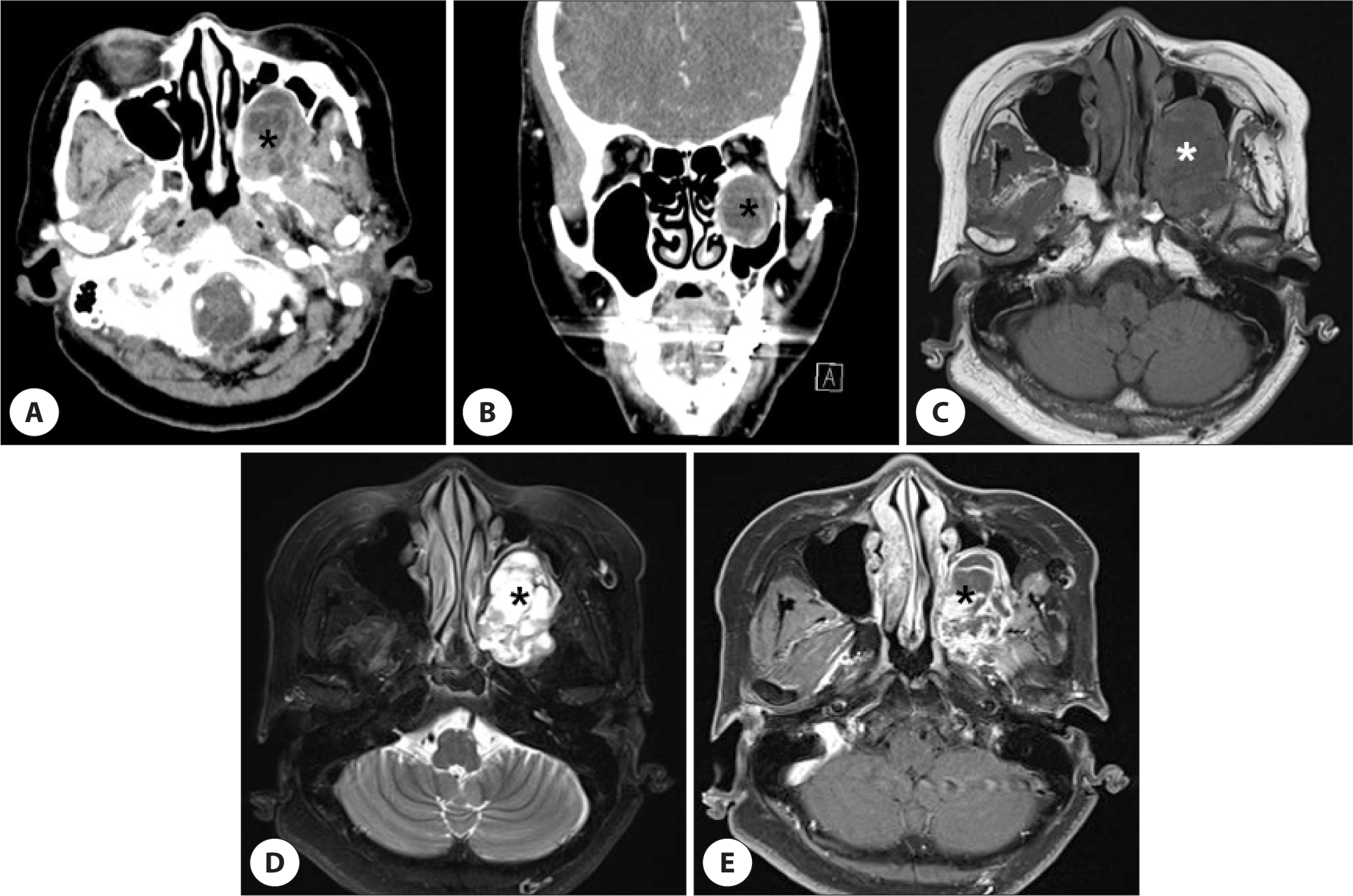
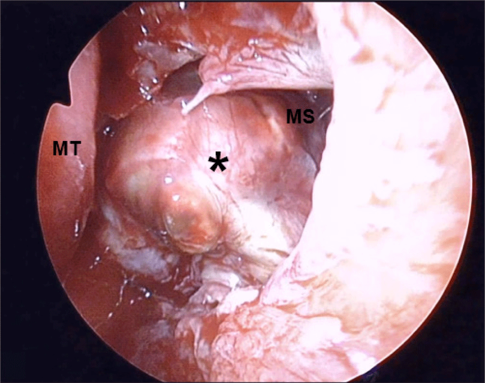
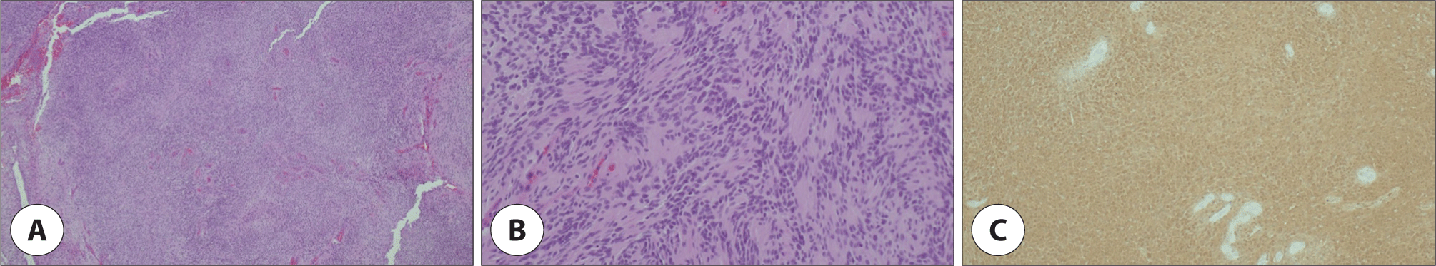
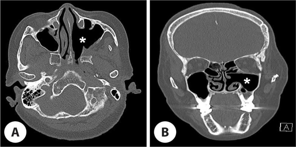
68세 여자 환자가 내원 1개월 전 건강검진 목적으로 타병원에서 촬영한 뇌 자기공명영상 검사상 좌측 익상구개와와 좌측 상악동의 측벽과 후벽 공간을 점유하는 종괴가 발견되어, 추가 검사 및 치료를 위해 본원에 내원하였다. 환자는 고혈압 외에는 과거력, 가족력 포함 특이소견이 없었다. 문진 및 안면 및 구강 내 촉진을 포함한 이학적 검사 상 이상소견이 없었고, 비인강경검사 소견상 좌측으로 완만한 비중격 만곡증 외 다른 특이소견은 없었다. 부비동 컴퓨터 단층촬영상 4.8×2.9×3.6 cm 크기의 비교적 경계가 분명한 불균질한 조영 증강되는 종괴가 좌측 익상구개와를 중심으로 좌측 상악동의 측벽과 후벽의 골파괴를 동반하여 점유하고 있는 소견을 보였고(Fig. 1A, B), 해당 병변은 부비동 자기공명영상 검사상 T1 강조영상에서 저신호 강도(Fig. 1C), T2 강조영상에서는 고신호 강도(Fig. 1D), gadolinium-T1 강조영상에서 조영증강(Fig. 1E) 소견을 보였다. 이를 토대로 신경원성 종양, 악성종양을 모두 염두해 두고, 비내시경 하 경상악동 접근법을 통한 절제 생검을 계획하였다. 수술은 전신마취하 우선적으로 비중격 만곡을 교정하기 위해 내시경하 비중격 교정술을 시행하였고, 그 후 좌측 중비도의 자연공을 넓히니 상악동 내에 비교적 피막으로 잘 둘러싸인 황색의 원형 종물(Fig. 2)이 관찰되었다. 최대한 피막을 보존하면서 미세 소파기(micro curette)를 이용하여 내부 점막과 분리하였고, 지라프 겸자(giraffe corceps)로 종양을 잡아 주변 조직과 분리하여 일부 우선 제거 후 동결절편 조직검사를 의뢰, 양성 신경인성 종양임을 확인하였다. 종양을 앞쪽으로 들어내니 상악동 후벽 및 측벽은 완전 소실된 상태였고, 익상구개와 공간으로 연결되어 있는 것이 확인 되었다. 익상구개와 공간내로부터 기시하는 종양을 제거하면서 내상악동맥이 손상을 받아 박동성 출혈이 있었으나, 전기적 소작술로 지혈하였으며, 적출한 검체는 조직검사 의뢰하였다. 조직병리 소견은 hematoxylin and eosin(H&E) 염색에서 세포 밀도가 높고 낮은 영역이 모두 존재하는 이상성 패턴의 책상배열 모양을 이루는 방추형의 신경세포 핵 증식이 관찰되었고(Fig. 3A, B), 면역조직화학 염색 200배에서 S-100 단백질 양성이 확인되어 신경초종으로 최종 진단되었다(Fig. 3C). 환자는 술 후 직후부터 안면부 따끔거리는 이상감각이 있었으나, 1개월 이후부터 완전 소실되었으며, 술 후 1년이 지난 현재 특별한 합병증 및 재발 소견 없이 외래 추적관찰 중이다(Fig. 4).
Fig. 1.
Preoperative computed tomography (CT) and magnetic resonance imaging (MRI) findings. Axial (A) and coronal (B) enhanced CT scans showed about 4.8×2.9×3.6 cm sized heterogenous enhancing mass lesions (black asterisk) on left pterygopalatine fossa with bony remodeling, focal bony defect and extending to left lateral and posterior maxillary sinus area. The mass was shown a low signal intensity in T1-weighted image (C, white asterisk), high signal intensity in T2-weighted image (D, black asterisk) and heterogenous signal intensity in T1-weighted gadolinium enhanced image (E, black asterisk).
Download Original Figure
Fig. 2.
Endoscopic endonasal intraoperative view of the tumor (black asterisk) during resection. MT: middle turbinate, MS: maxillary sinus.
Download Original Figure
Fig. 3.
The histopathologic findings. A: Spindle cell proliferative lesion with thickened blood vessels (H&E, ×40). B: It shows that biphasic pattern (hypercellular and hypocellular area), nuclear palisading (H&E, ×200). C: The neoplastic cells show uniform cytoplasmic expression of S-100 protein (Immunohistochemical staining for S-100, ×200).
Download Original Figure
Fig. 4.
Postoperative computed tomography (CT) findings. Axial (A) and coronal (B) non-enhanced CT scans showed total excision of the tumor and clearance of left pterygopalatine fossa (white asterisk).
Download Original Figure
고찰
신경초종은 신경섬유의 신경초가 있는 뇌신경, 교감신경 및 말초신경이 있는 신체의 어느 부위에서나 발생할 수 있고,4) 전체 신경초종 중 두경부 영역에서는 발생하는 빈도는 25%–45%이며, 이중 가장 흔한 위치는 제8번 뇌신경(80%)으로, 그 외 목(8%), 이하선(7%), 볼(7%), 혀(6%), 두피(6%), 부비강(3%–4%) 순의 빈도를 나타낸다.3,5,6) 임상적으로 종양의 기원은 비강이나 부비동에서 발생하는 경우 삼차신경의 안분지나 상악분지, 자율신경이 흔하고, 부인강에 발생한 경우 대개 미주신경과 경부 교감 신경절이 흔하다.5,7)
신경초종의 임상 증상은 초기에는 무증상으로 무통성 종괴 양상을 보이나, 종물의 크기가 증가함에 따라 해부학적 위치에 따른 주변 구조물을 압박하여 증세가 발현된다.1) 익상구개와에서 기시하는 경우, 비강 쪽으로 침범 시 편측성 비폐색, 비출혈, 비루, 안면부종, 안면통증 및 감각이상, 후각장애 등의 다양하고 비특이적인 증상을 호소할 수 있고,1,2,4) 안와나 두개내로 침범시 안구운동 장애, 안구돌출, 안구통, 복시 등의 증상,1) 비인두의 경우 구강호흡, 코골이, 경부 이물감, 연하 곤란 등이 발생할 수 있다.7)
신경초종의 진단은 병력 및 임상증상, 영상검사 등을 바탕으로 면역조직화학 검사를 포함하는 조직병리학적 소견을 통해 최종 확진이 이루어진다.7–9)
신경초종의 방사선학적 소견은 비특이적인 경우가 흔하나, 종괴의 크기가 클수록 낭성변화, 출혈, 석회화 등으로 인해 더 불균질한 영상 소견을 보인다.10) 자기공명 영상은 T1 강조 영상에서 중등도, T2 강조 영상에서 전반적인 중등도, 부분적 이형 고신호 강도를 보인다.1,11)
신경초종의 조직병리소견은 안토니 A와 B 두 가지 소견이 있다.6,8,9) 안토니 A는 핵들이 울타리모양을 이뤄 형성된 verocay body가 관찰되는 특징적인 반면, 안토니 B는 느슨하게 배열된 망상 조직에서 다양한 형태의 핵모양이 나타난다.8–10) 또한 조직병리학적 소견에 따라 세포성과 비세소성으로 구분 짓는데, 세포성 신경초종의 경우 안토니 A의 영역이 현저히 우세하고, verocay body가 없는 것이 특징적이다. 반면 두 가지 모두 S-100 단백질에 대한 면역화학염색에서 양성소견을 갖는다.10)
신경초종의 치료는 방사선 치료에 저항성이 있어, 원발 종양의 광범위 절제를 원칙으로 하나, 종괴의 피막 일부가 남아있어도 재발은 잘 하지 않는다는 것으로 알려져 있다.4,9) 병변의 크기와 위치, 주변구조물에 따라 비내시경 수술, Caldewell-Luc 접근법, 외비절개, 측두하와 접근법, 구순하 접근법, 감마나이프 등을 통해 제거할 수 있으나 비내시경 수술이 가장 적합한 방법으로 알려져 있다.9,12) 익상구개와는 익상구개 신경절, 익돌관 신경, 제5신경의 상악분절, 내상악동맥의 말단 분지, 지방 등으로 구성되어 있다.13) 본 증례는 무증상의 우연히 발견된 익상구개와에서 기시하여 상악동을 점유한 신경초종의 경우로 수술적 시야 확보와 술후 합병증, 미용적 측면을 고려하였을 때 비내시경을 통한 경상악동 접근법으로 안전하게 제거가 가능하다는 것에 의미를 두며, 이러한 최소 침습적인 방법을 적극적으로 동원해 치료하는 것이 타당하다는 교훈을 얻을 수 있었다.