Case Report

편도절제술 이후 동시에 진단된 두 종류의 림프종 1예

이승주1https://orcid.org/0000-0001-8276-2567, 박근우1https://orcid.org/0000-0002-9187-4381, 박체현1https://orcid.org/0009-0007-7102-8851, 정태영1,*https://orcid.org/0000-0001-9029-2075
Seung Joo Lee1https://orcid.org/0000-0001-8276-2567, Geun Woo Park1https://orcid.org/0000-0002-9187-4381, Che Hyun Park1https://orcid.org/0009-0007-7102-8851, Tae Young Jung1,*https://orcid.org/0000-0001-9029-2075
Author Information & Copyright
1메리놀병원 이비인후과
1Departments of Otorhinolaryngology - Head and Neck Surgery, Maryknoll Hospital, Busan, Korea
*Corresponding author: Tae Young Jung, Department of Otorhinolaryngology-Head and Neck Surgery, Maryknoll Hospital, Busan 48972, Korea, Tel: +82-51-461-2692, Fax: +82-51-462-9419, E-mail: handsjung@naver.com

© Copyright 2023 The Busan, Ulsan, Gyeoungnam Branch of Korean Society of Otolaryngology-Head and Neck Surgery. This is an Open-Access article distributed under the terms of the Creative Commons Attribution Non-Commercial License (http://creativecommons.org/licenses/by-nc/4.0/) which permits unrestricted non-commercial use, distribution, and reproduction in any medium, provided the original work is properly cited.

Received: Oct 11, 2023; Revised: Nov 08, 2023; Accepted: Nov 21, 2023

Published Online: Dec 31, 2023

ABSTRACT

Lymphoma is a malignant tumor that occurs in the immune system and is largely classified into Hodgkin’s lymphoma (HL) and non-Hodgkin’s lymphoma (NHL). Follicular lymphoma, one subtype of NHL, can histologically transform into more aggressive cancers due to the nature of the disease: diffuse large B cell lymphoma (DLBCL), Burkitt lymphoma, and lymphoblastic lymphoma. We present a case of two different subtypes of lymphoma diagnosed simultaneously after a tonsillectomy in a 37-year-old female: DLBCL and follicular lymphoma.

Keywords: Tonsillectomy; Lymphoma, follicular; Lymphoma, large B-cell, diffuse

서론

림프종은 면역 기관에서 생기는 악성 종양으로 무통성에서부터 공격적인 종양의 집합체를 의미하며 크게 호치킨스 림프종(Hodgkin’s lymphoma, HL), 비호치킨스 림프종(non-Hodgkin’s lymphoma, NHL)로 분류한다. 그 중 비호치킨스 림프종은 대부분 일차적으로 림프절에서 발생하지만 구인두, 골수, 장, 피부 등 20%–30%에서 림프절 외(extranodal)에서의 발생이 보고되어 있다.1) 그 중 구강에서의 발생은 드물며, 보고된 림프종 환자의 3%–5%에서 확인되며, 두경부 부위에서는 림프절 외에는 왈데이어환(Waldeyer’s ring)에서 가장 흔히 발생한다.2)

구개 편도에 림프종이 발생하는 경우는 흔히 보고되나, 양쪽 편도에서 각각 다른 림프종이 확인된 경우는 국내에서 보고된 바가 없다. 이에 본 저자들은 일측 편도 덩이를 주소로 양측 편도 절제술을 시행하여, 좌측에 소포림프종(follicular lymphoma), 우측에 미만성 거대 B세포 림프종(diffuse large B cell lymphoma, DLBCL)으로 서로 다른 림프종이 진단된 1례를 경험하였기에 이를 보고하는 바이다.

증례

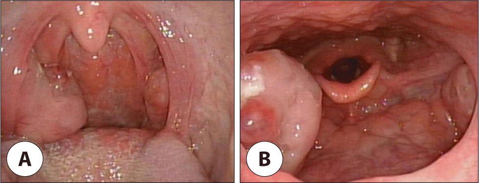
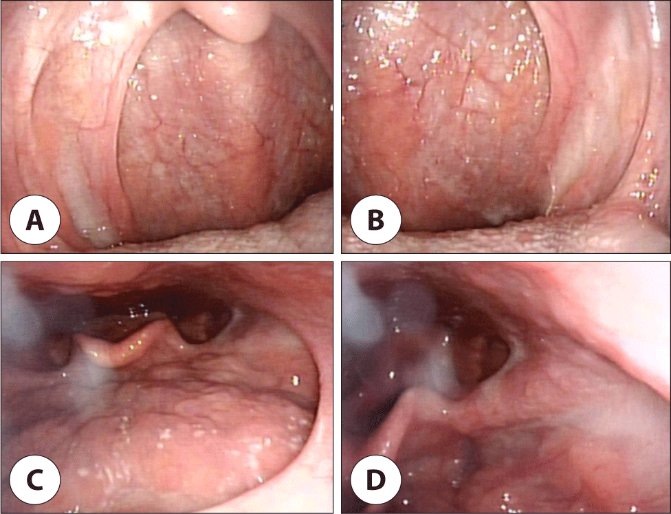
37세 여자가 4개월 전부터 생긴 목 이물감으로 개인병원에서 치료받던 중 호전이 없어 본원 외래에 내원하였다. 내과적 기저 질환은 없으며, 환자는 과거 연 3회 정도의 편도염 과거력 있었고, 내원 당시 연하 불편감 및 간헐적인 호흡 불편감을 호소하였다. 이학적 검사상 목 주변으로 만져지는 종물은 없었고, 내시경 상 편도는 프리드만(Friedman) 분류에 따라 우측은 3단계, 좌측은 1단계 크기의 편도가 확인되었다(Fig. 1). 수술 전 피검사나 흉부 X선 검사 상 이상소견은 관찰되지 않았다. 저자들은 악성 종물 가능성 배제할 수 없고 과거 편도염의 과거력 있어 양측 편도 절제술을 시행하였다.

jcohns-34-4-215-g1
Fig. 1. The pre-operative endoscopic view of tonsil size evaluated by Frideman classification is Grade 1, 3. A: Oral cavity view. B: Laryngoscopic view.
Download Original Figure

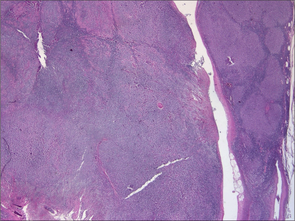
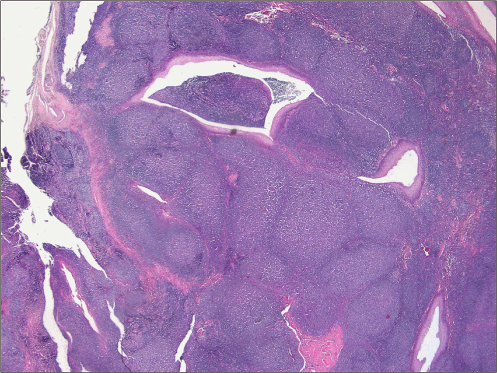
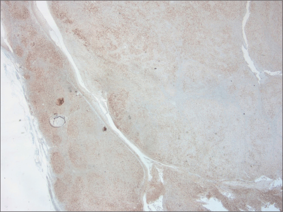
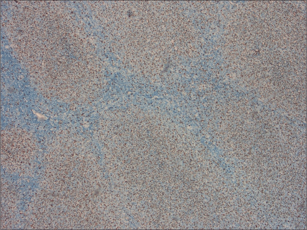
수술 2일째 특이 소견 없이 퇴원하였고, 수술 후 13일째 방문한 외래에서는 목 통증 일부 호소하였으나 수술 부위에는 이상이 없었다. 조직검사 상 우측 편도는 미만성 거대 B세포 림프종에 동반된 미만성 패턴(25% 이하)(Fig. 2), 좌측은 소포림프종으로 확인되었다(Fig. 3). 아종(subtype)을 확인하기 위해 시행한 면역형광염색에서 좌측은 CD20 양성, CD3 양성, 우측은 Bcl-6 양성(Fig. 4), CD10 음성, Bcl-2 음성, Ki67 50% 이상으로(Fig. 5) 좌측 소포림프종, 우측 미만성 거대 B세포 림프종에 해당하는 소견을 보였다.

jcohns-34-4-215-g2
Fig. 2. Tumor shows diffuse area (Lt. side) & preserved follicular pattern (Rt. Side) of neoplastic cells (H&E stain, ×10).
Download Original Figure
jcohns-34-4-215-g3
Fig. 3. The neoplastic follicles are closely packed with lacking mantle zones (H&E stain, ×10).
Download Original Figure
jcohns-34-4-215-g4
Fig. 4. The germinal center cells are positive for Bcl-6 (Immunohistochemical stain for Bcl-6, ×10).
Download Original Figure
jcohns-34-4-215-g5
Fig. 5. The tumor shows more than 50% of Ki67 labelling index (Immunohistochemical stain for Ki67, ×40).
Download Original Figure

구인두 림프종 진단 하 항암치료 시행 위해 혈액종양내과로 의뢰하였고 Ann arbor stage에 따라 ileocecal, mesentery lymph node에 침범되어 있고, 발열, 야간 발한, 체중 감소와 같은 systemic B symptom이 없어 stage IIIA로 진단되었다. 6차까지의 R-CHOP(rituximab, cyclophosphamide, doxorubicin, vincristine, prednisone) 항암치료 시행하였다. 현재 완전 관해(complete remission) 상태이며, 이비인후과 및 혈액종양내과에서 별다른 재발 없이 경과 관찰 중이다(Fig. 6).

jcohns-34-4-215-g6
Fig. 6. The post-operative endoscopic view 10 months after surgery. A, B: Oral cavity view. C, D: Laryngoscopic view.
Download Original Figure

고찰

소포림프종은 저분화 B세포 림프종 중 2번째로 흔하며 성인의 비호치킨 림프종의 약 30%에서 발생한다. 또한 임상증상이 대부분 무통성(indolent)이며 생존률이 높지만, 현재 완치는 불가능하다.1) 2017년 WHO 분류에 따르면 고배율(high power field, HPF) 하에서 중심아세포(centroblast)의 비율에 따라 크게 Grade 1, 2, 3으로 분류한다: Grade 1: 0–5개/HPF, Grade 2: 6–15개/HPF, Grade 3: >15개/HPF. 이중 Grade 3는 다시 중심세포(centrocyte)가 확인될 시 3A, 교형 판상(solid sheet)의 중심아세포가 확인될 시 3B로 분류한다. 3A의 경우, 형질 전환의 위험성이 증가된 것으로 알려져 있으며,3) Grade 3B의 경우, 새로 발생한 공격적인 림프종과 매우 관련이 깊은 것으로 알려져 있다.4) 이로 인해 Grade 3는 Grade 1, 2와 달리 미만성 거대 B세포 림프종에 유사하게 치료한다.5) 소포림프종의 특징은 14번과 18번 염색체의 전위(translocation)로 생기는 BCL-2 유전자의 과발현이며, 85%에서 이를 확인할 수 있다.6) 면역염색 시 CD19, CD20, CD10, Bcl-6에서 양성을 보이며, CD5, CD23에서 음성을 보인다.

미만성 거대 B세포 림프종은 다양한 스펙트럼을 가진 림프구성 종양의 집합체로써 종종 야간 발한, 체중감소, 발열과 같은 전신 증상(B symptom)을 동반한다.1) 전신 증상을 포함하여 공격적이나 5년 생존률이 80%–85%에 달하여 치료에 반응이 좋은 것이 특징이다.7) CD10은 배중심에 존재하는 막성 금속단백가수분해효소의 표지자로 대부분의 소포림프종에서 발현하며, 미만성 거대 B세포 림프종에서는 20%–40%에서 발현한다. CD3는 T-세포에서 발현하며 세포질에 신호전달을 담당하는 표지자로 발암화와 연관되어 있다.1) Ki67의 경우, 세포분열과 관련된 마커로 조직학적 변형이 일어난 림프종에서는 높게 발현하는 것이 특징이다.8) Bcl-6는 정상 림프 조직의 배중심 B세포(Germial B cell)에서 발현하며 미만성 거대 B세포 림프종에서는 57%–100%에서 발현된다.9)

본 증례에서 좌측 편도에 소포림프종의 면역염색결과상 Grade 2, 면역염색 결과상 CD20 양성, CD3 양성으로 확인되었으며, 우측 편도에 미만성 거대 B세포 림프종에서는 CD20 양성, CD3 양성, Bcl-6 양성, CD10 음성, Bcl-2 음성, Ki67 50% 이상으로 확인되었다.

소포림프종은 병의 특성상 더 공격적인 암으로 조직학적 변형(histological transformation)이 일어날 수 있다. 가장 흔한 변형은 미만성 거대 B세포 림프종으로의 변형(30%–40%)이며, 그 외에도 버킷 림프종, 림프모구 림프종, 때때로는 호치킨 림프종으로의 변형이 알려져 있다.10) 소포림프종의 미만성 거대 B세포 림프종으로의 조직학적 변형은 매년 3% 정도 일어나며, 약 16년 정도 지난 이후에는 더 증가하지 않고 고점을 이루며 10%–60%의 비율로 전환될 수 있다.11,12) 조직학적 변형은 여러 유전자의 변형으로 생기며 많은 연구가 이루어지고 있다. 주로 알려진 조직학적 변형은 더블 히트(double hit) 기전으로 BCL-2 유전자의 전위가 이미 일어난 세포에서 MYC 유전자의 활성화가 일어남으로 더욱 조직학적 변형이 잘 되는 세포로 변하는 것으로 알려져 있다.3) 이와 더불어 트리플 히트(triple hit) 기전으로 더블 히트 기전에서 추가로 BCL-6 유전자의 전위가 일어난 림프종은 더욱 나쁜 예후와 관련이 있다.13) 추가적으로 세포주기와 DNA 손상복구에 관련되어 CDKN2A/B의 소실을 확인할 수 있는데, 이는 DNA 복구와 관련된 p53 gene의 비활성화, 세포주기의 조절과 관련된 p16의 과발현이 특징적이다.14,15)

본 증례에서는 크기가 크지 않고 모양도 보통의 편도와 크게 다르지 않았던 좌측 편도에서도 소포림프종이 확인되었다. 일측의 병변이 악성이 의심될 경우, 반대편의 정상 조직처럼 보이는 부위도 조직검사를 시행하고 결과를 확인하는 것이 필요하다.

결론

림프종 내에서도 소포림프종과 같은 저위험군 림프종에서 미만성 거대 B세포 림프종과 같은 공격적인 암으로 조직학적 변형이 일어날 수 있음을 기억하고 정상적으로 보이는 조직에서도 림프종의 가능성을 의심해봐야 되겠다.

Acknowledgements

Not applicable.

Funding Information

Not applicable.

Conflicts of Interest

No potential conflict of interest relevant to this article was reported.

Author Contribution

Conceptualization: Jung TY.

Data curation: Park GW.

Formal analysis: Lee SJ.

Methodology: Lee SJ, Jung TY.

Software: Park CH.

Validation: Jung TY.

Investigation: Lee SJ.

Writing - original draft: Lee SJ.

Writing - review & editing: Lee SJ, Park GW, Park CH, Jung TY.

Ethics Approval

Not applicable.

References

1.

Mittal M, Puri A, Nangia R, Sachdeva A. Follicular lymphoma transforming into anaplastic diffuse large B-cell lymphoma of oral cavity: a case report with review of literature. J Oral Maxillofac Pathol 2015;19(3): 379-84.

2.

Jacobs C, Hoppe RT. Non-Hodgkin’s lymphomas of head and neck extranodal sites. Int J Radiat Oncol Biol Phys 1985;11(2):357-64.

3.

Kridel R, Mottok A, Farinha P, Ben-Neriah S, Ennishi D, Zheng Y, et al. Cell of origin of transformed follicular lymphoma. Blood 2015;126(18):2118-27.

4.

Swerdlow SH, Campo E, Pileri SA, Harris NL, Stein H, Siebert R, et al. The 2016 revision of the World Health Organization classification of lymphoid neoplasms. Blood 2016;127(20):2375-90.

5.

Shankland KR, Armitage JO, Hancock BW. Non-Hodgkin lymphoma. Lancet 2012;380(9844):848-57.

6.

Campo E, Swerdlow SH, Harris NL, Pileri S, Stein H, Jaffe ES. The 2008 WHO classification of lymphoid neoplasms and beyond: evolving concepts and practical applications. Blood 2011;117(19):5019-32.

7.

Martelli M, Ferreri AJM, Agostinelli C, Di Rocco A, Pfreundschuh M, Pileri SA. Diffuse large B-cell lymphoma. Crit Rev Oncol Hematol 2013;87(2):146-71.

8.

Natkunam Y, Soslow R, Matolcsy A, Dolezal M, Bhargava V, Knowles DM, et al. Immunophenotypic and genotypic characterization of progression in follicular lymphomas. Appl Immunohistochem Mol Morphol 2004;12(2):97-104.

9.

Bhattacharyya I, Chehal HK, Cohen DM, Al-Quran SZ. Primary diffuse large B-cell lymphoma of the oral cavity: germinal center classification. Head Neck Pathol 2010;4(3):181-91.

10.

Fischer T, Zing NPC, Chiattone CS, Federico M, Luminari S. Transformed follicular lymphoma. Ann Hematol 2018;97(1):17-29.

11.

Takata K, Miyata-Takata T, Sato Y, Yoshino T. Pathology of follicular lymphoma. J Clin Exp Hematopathol 2014;54(1):3-9.

12.

Lossos IS, Gascoyne RD. Transformation of follicular lymphoma. Best Pract Res Clin Haematol 2011; 24(2):147-63.

13.

Akasaka T, Lossos IS, Levy R. BCL6 gene translocation in follicular lymphoma: a harbinger of eventual transformation to diffuse aggressive lymphoma. Blood 2003;102(4):1443-48.

14.

Pasqualucci L, Khiabanian H, Fangazio M, Vasishtha M, Messina M, Holmes AB, et al. Genetics of follicular lymphoma transformation. Cell Rep 2014;6(1):130-40.

15.

Sherry MA, Best TM. A comparison of 2 rehabilitation programs in the treatment of acute hamstring strains. J Orthop Sports Phys Ther 2004;34(3):116-25.