Original Article

폐쇄성 수면 무호흡 환자의 후두개 폐쇄 양상

지영훈1https://orcid.org/0000-0003-4994-6254, 우주영1https://orcid.org/0000-0002-5972-5714, 고태경1https://orcid.org/0000-0002-2805-2270, 구수권1,*https://orcid.org/0000-0002-7541-1985
Young Hoon Chi1https://orcid.org/0000-0003-4994-6254, Joo Young Woo1https://orcid.org/0000-0002-5972-5714, Tae Kyung Koh1https://orcid.org/0000-0002-2805-2270, Soo Kweon Koo1,*https://orcid.org/0000-0002-7541-1985
Author Information & Copyright
1부산성모병원 이비인후과
1Department of Otorhinolaryngology-Head and Neck Surgery, Busan Saint Mary’s Hospital, Busan, Korea
*Corresponding author: Soo Kweon Koo, Department of Otolaryngology-Head and Neck Surgery, Busan Saint Mary’s Hospital, Busan 48575, Korea, Tel: +82-51-933-7214, Fax: +82-51-956-1956, E-mail: koosookweon@naver.com

© Copyright 2023 The Busan, Ulsan, Gyeoungnam Branch of Korean Society of Otolaryngology-Head and Neck Surgery. This is an Open-Access article distributed under the terms of the Creative Commons Attribution Non-Commercial License (http://creativecommons.org/licenses/by-nc/4.0/) which permits unrestricted non-commercial use, distribution, and reproduction in any medium, provided the original work is properly cited.

Received: Aug 11, 2023; Revised: Sep 28, 2023; Accepted: Oct 20, 2023

Published Online: Dec 31, 2023

ABSTRACT

Background and Objectives:

We evaluated the pattern of epiglottic obstruction in obstructive sleep apnea (OSA) patients, focusing on the results of drug induced sleep endoscopy (DISE), along with a literature review.

Materials and Methods:

The subjects were 270 Koreans who underwent DISE. Each patient underwent a thorough history taking, physical examination, polysomnography and DISE image evaluated by one otolaryngologist.

Results:

The prevalence of epiglottic obstruction was 39% (105/270), anterior-posterior complete obstruction was 26% (70/270), and partial obstruction was 13% (35/270). There was no significant correlation between gender, age, body mass index and epiglottis obstruction. The more severe the epiglottic obstruction, the more severe the sleep apnea. The more severe the anterior-posterior obstruction of the tongue base, the more severe the obstruction of the epiglottis (p<0.001).

Conclusion:

The epiglottis is not well understood compared to other anatomical sites, and it can OSA either alone or simultaneously with other obstructive sites, so researches are needed from a therapeutic point of view.

Keywords: Sleep apnea; obstructive; Epiglottis; Polysomnography

서론

폐쇄성 수면 무호홉(obstructive sleep apnea, OSA)의 유병율은 일반 성인 인구의 9%–38% 정도이며, 여성보다는 남성에서 그리고 나이가 들수록 OSA의 유병율은 증가하는 것으로 알려져 있다.1) OSA는 과도한 주간 졸림, 심혈관계 질환, 인지장애, 각종 대사장애 등에 많은 영향을 미치고, 이는 환자의 사망률을 높이는 원인으로 정확한 진단과 치료가 중요하다.2) 지속적인 양압요법(continuous positive airway pressure, CPAP)은 OSA 환자에서 가장 먼저 고려되는 치료이다. 하지만 장기간 사용에 따른 순응도 문제로 외과적 치료가 고려되기도 하는데,3,4) 코골이와 OSA의 수술적 치료계획을 위해서는 폐쇄 부위, 폐쇄 형태, 폐쇄에 관여하는 해부학적 구조를 찾는 것이 중요하다. 그동안 환자의 폐쇄 부위를 찾는 여러 가지 검사가 개발되었고, 수면상태에서 정확한 진단을 위한 노력들이 있었다. CT, MRI(magnetic resonance imaging), cephalometry 등은 각성 시 상기도를 비교적 잘 보여주지만 정적이며 수면상태에서의 동적인 상기도를 진단하는 데는 한계가 있다. 1991년 Croft와 Pringle이 약물로 수면을 유도한 후 내시경을 통해 관찰하는 “sleep nasoendoscopy”를 제안한 후,5) 약물 유도 수면 내시경(drug induced sleep endoscopy, DISE)은 수면 상태에서 폐쇄 부위를 진단하는 새로운 진단 기법으로, 현재 전세계적으로 널리 사용되고 있다. DISE는 OSA의 병태생리에 대한 이해를 향상시켰을 뿐 아니라, 수면 시 폐쇄 부위를 진단함으로써 치료적인 측면에서 많은 도움을 주고 있다. OSA 환자의 폐쇄 부위는 연구개, 구인두, 혀뿌리, 및 후두개 등으로 다양하며, 대개 여러 가지 구조가 복합적인 폐쇄를 보이지만 단독으로 폐쇄가 일어나기도 한다. 현재까지 OSA 환자의 폐쇄 부위와 관련된 많은 연구가 진행되었지만 폐쇄가 일어나는 기전이나 질환과의 연관성에 대해서는 아직도 의문점이 많다. 특히 후두개 폐쇄는 OSA 환자의 일차 치료인 CPAP 치료의 장애가 될 수 있는데, 이는 후두개가 해부학적으로 CPAP의 공기 흐름의 반대편에 위치하여 후두개 폐쇄를 악화시킬 수 있기 때문이며, 이런 관점에서 후두개 폐쇄의 관심이 높아지고 있다. 저자들은 OSA 환자들에게 시행한 DISE의 결과를 중심으로 후두개의 폐쇄의 형태를 문헌고찰과 함께 연구하였다.

대상 및 방법

연구의 일반적인 설정

본원 이비인후과를 내원하여 DISE를 시행한 한국인 270명을 대상으로 하였다(Table 1). 남자 232명, 여자 38명으로 평균연령은 42.85±12.33, 평균 BMI(body mass index)는 26.42±3.72였으며, 평균 AHI(anpea-hypopnea index)는 32.01±22.75였다. 검사자 간의 오차를 배제하기 위하여 동일한 한 명의 검사자가 시행하였으며, 후향적 연구로 본원 IRB의 심사를 거쳐 승인되었다. DISE 이전에 모든 환자는 철저한 귀, 코, 목 검사와 병력을 청취하였으며, 명백한 retrognathia를 가진 환자, 하악 이형성증, 또는 이전에 연구개 또는 혀의 수술을 받은 사람은 제외되었다. 각 환자는 철저한 병력 청취, 신체 검사, 수면다원검사(polysomnography, PSG) 및 DISE를 시행하였다. 수면다원검사는 full PSG 시스템(EMBLA Embletta MPR-PG, Flaga, Iceland)을 이용하여 AASM 지침에 따라 scoring하였다.

Table 1. Demography of the patients (n=270)
Characteristics Value
Male 232
Female 38
Mean age±S.D. (yr) 42.85±12.33
Mean BMI±S.D. (kg/m2) 26.42±3.72
AHI 32.01±22.75
Supine AHI 49.37±55.97
Lateral AHI 16.18±22.49

Data are presented as n or mean±S.D.

BMI: body mass index, AHI: anpea-hypopnea index.

Download Excel Table
Drug induced sleep endoscopy (DISE)

모든 DISE는 같은 이비인후과 전문의가 조용하고 약간 어두운 수술실에서 시행하였다. 마취과 의사의 도움을 받아 호흡 모니터링 하에서 시행하였으며, 앙와위 자세의 환자에게 midazolam을 정맥 주사하여 수면을 유도했다. Midazolam은 환자당 0.07 mg/kg으로 천천히 주사하였으며, 목표 제어 주입 시스템(target-controlled infusion system)을 사용하여 한번에 1–2.5 mg, 최대 7.5 mg을 주입하였다. 환자가 수면이 유도되면 청각 반응을 관찰하여 진정 정도(Ramsay의 진정 정도 5)가 일정 수준에 도달하면 직경 4 mm의 유연 비디오 후두내시경(flexible videolaryngoscope)을 코를 통해 부드럽게 삽입하여 상기도의 폐쇄를 관찰하고 녹화하였으며, 이 영상은 한 사람의 이비인후과 의사가 분석하였다.6)

분류 체계

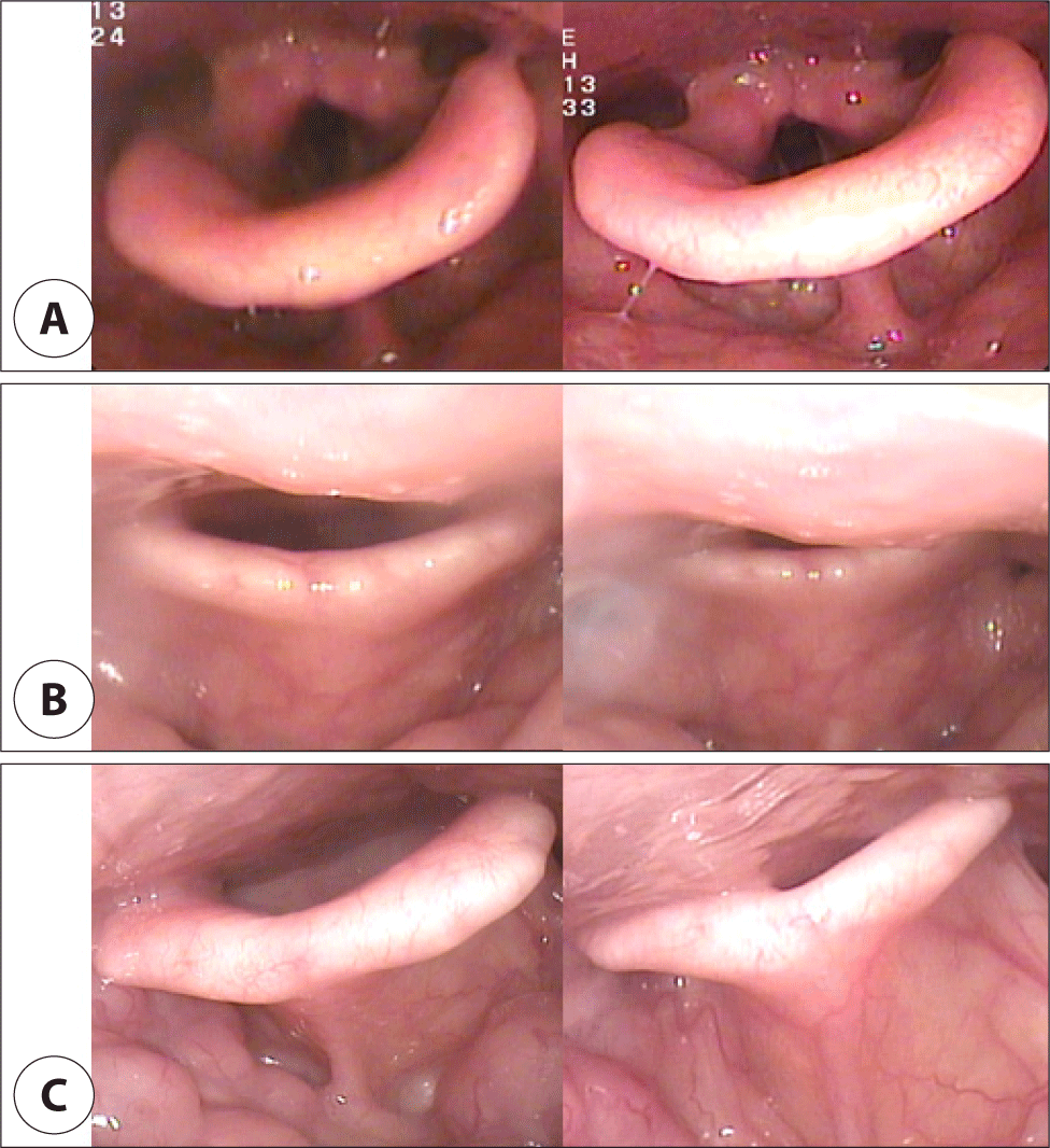
저자들은 기도를 연구개 후방 부위(retropalatal level)와 혀 후방 부위(retrolingual level)로 나누고, 이를 바탕으로 수정된 VOTE 분류를 만들었다.7,8) 저자들의 분류 체계는 폐쇄 부위의 위치, 정도 및 원인이 되는 해부학적 구조를 포함했다. 연구개 후방 부위는 연구개(전후 직경), 측면 인두벽(외측 직경), 편도선(폐쇄에 기여하는 특정 구조)으로 세분화하였다(Fig. 1). 혀 후방 부위는 혀뿌리(전후 직경), 측면 인두벽(외측 직경), 후두개(폐쇄에 기여하는 특정 구조)로 분류하였다. 기도 폐쇄 정도는 폐쇄 없음(0), 부분 폐쇄(1, 50%–75%)와 완전 폐쇄(2, >75%)로 분류하였다(Table 2).

jcohns-34-4-136-g1
Fig. 1. Representative photos according to the degree of epiglottis obstruction. A: Patient with degree of obstruction 0. B: Patient with degree of obstruction 1. C: Patient with degree of obstruction
Download Original Figure
Table 2. Classification of DISE findings
Level Degree of obstruction* Configuration
AP diameter Lat. diameter Contributing structure
Retropalatal 0/1/2 Palate LPW Tonsil
+/– +/– +/–
Retrolingual 0/1/2 Tongue base LPW Epiglottis
+/– +/– +/–

* Degree of obstruction has one number of each structure: 0=no obstruction (no vibration), 1=partial obstruction (vibration, 50%–75%), 2=complete obstruction (collapse, >75%).

Dichotomous configuration noted for structures with degree of obstruction greater than 0.

DISE: drug-induced sleep endoscopy, AP: antero-posterior, Lat.: lateral, LPW: lateral pharyngeal wall.

Download Excel Table
통계 분석

연구 대상자의 일반적 특성과 후두개 폐쇄와의 상관관계를 알아보기 위하여 교차분석을 시행하였으며, 연령과의 상관관계를 알아보기 위해 일원배치분산분석을 시행하였다. DISE에서 관찰되는 여러 폐쇄 부위와 후두개 폐쇄와의 상관관계는 교차분석을 시행하였다.

결과

후두개 폐쇄 유병율

후두개 폐쇄 유병율은 39%(105/270)였고, 전후방 완전 폐쇄가 26%(70/270), 부분 폐쇄가 13%(35/270)였다. 모두 여러 군데 동시 폐쇄가 일어난 경우였고 후두개 단독 폐쇄는 없었다.

DISE(drug induced sleep endoscopy)에서 성별, 연령, BMI(body mass index)와 후두개 폐쇄와의 연관성(Table 3)
Table 3. Correlation between gender, age, BMI and epiglottic obstruction in DISE (n=270)
Variable Level Epiglottic obstruction (n (%)) χ2/F-value p-value
0 (n=166) 1 (n=35) 2 (n=70)
Sex Male (%) 142 (61.2) 29 (12.5) 61 (26.3) .36 .835
Female (%) 23 (60.5) 6 (15.8) 9 (23.7)
Age±S.D. (yr) 42.90±12.38 44.06±11.85 42.11±12.54 .29 .747
BMI±S.D. (kg/m2) 26.21±3.52 26.46±4.18 26.87±3.95 .78 .461
 <25 72 (64.9) 15 (13.5) 24 (21.6) 2.66 .617
 25–30 71 (60.2) 13 (11.0) 34 (28.8)
 ≥30 23 (54.8) 7 (16.7) 12 (28.6)

Data are presented as n (%) or mean±S.D.

BMI: body mass index, DISE: drug-induced sleep endoscopy.

Download Excel Table

DISE에서 성별, 연령, BMI와 후두개 폐쇄와의 연관성을 통계적 분석한 결과, 유의미한 상관관계를 나타내지 않았다. 연령의 경우, 10세 단위로 구간을 나누어도 유의미한 결과는 확인되지 않았으며, BMI는 연속형 변수로 고려하여도 역시 유의미한 차이는 없었다.

DISE(drug induced sleep endoscopy)에서 수면다원검사 결과와 후두개 폐쇄의 연관성(Table 4)
Table 4. Correlation between polysomnographic results and epiglottis obstruction in DISE
Variable Epiglottic obstruction F-value p-value Post Hoc
0 (n=166) 1 (n=35) 2 (n=70)
AHI 28.27±18.78 29.88±21.74 40.50±27.99 5.15 .007 0,1<2

Data are presented as mean±S.D.

DISE: drug-induced sleep endoscopy, AHI: anpea-hypopnea index.

Download Excel Table

DISE에서 수면다원검사 결과와 후두개 폐쇄의 연관성에 대해 통계적으로 분석한 결과, 후두개 폐쇄가 심할수록 AHI가 높은 것으로 나타났다.

DISE(drug induced sleep endoscopy)에서 후두개 폐쇄와 다른 구조 폐쇄와의 연관성(Table 5)
Table 5. Correlation between epiglottis obstruction and other structural obstruction in DISE
Variable Level Epiglottic obstruction (n (%)) χ2-value p-value
0 (n=166) 1 (n=35) 2 (n=70)
Palate AP 0 14 (53.8) 5 (19.2) 7 (26.9) 12.56 .014
1 21 (95.5) 0 ( 0.0) 1 ( 4.5)
2 131 (58.7) 30 (13.5) 62 (27.8)
Palate Lat 0 28 (56.0) 7 (14.0) 15 (30.0) 1.363 .851
1 30 (58.8) 8 (15.7) 13 (25.5)
2 108 (63.5) 20 (11.8) 42 (24.7)
Tonsil 0 108 (70.6) 12 ( 7.8) 33 (21.6) 24.78 <.001
1 15 (31.9) 13 (27.7) 19 (40.4)
2 43 (60.6) 10 (14.1) 18 (25.4)
TB AP 0 63 (84.0) 2 ( 2.7) 10 (13.3) 34.41 <.001
1 59 (62.1) 17 (17.9) 19 (20.0)
2 44 (43.6) 16 (15.8) 41 (40.6)
TB Lat 0 49 (62.0) 9 (11.4) 21 (26.6) 5.470 .242
1 69 (68.3) 10 ( 9.9) 22 (21.8)
2 48 (52.7) 16 (17.6) 27 (29.7)

Data are presented as n (%).

DISE: drug-induced sleep endoscopy, AP: anteroposterior, Lat: lateral, TB: tongue base.

Download Excel Table

DISE에서 후두개 폐쇄와 다른 구조 폐쇄와의 상관관계를 알아보기 위해 통계적으로 분석한 결과, 후두개의 완전 폐쇄(2, >75%)가 있는 경우, 혀뿌리 부분의 전후방 폐쇄 정도에 영향을 받은 것으로 나타났다(p<0.001).

고찰

DISE는 수면 중 기도의 폐쇄 부위를 진단하는데 유용한 검사법으로, 현재까지 한국인을 대상으로 진행한 연구는 많지 않았다. 본 연구에서는 이전 연구에서 저자들이 제안한 수정된 VOTE 분류를 사용하여 본원을 내원한 한국인을 대상으로 DISE를 시행하였다.

후두개 폐쇄는 그 방향과 정도에 따라 전후방, 측방, 부분적 폐쇄와 완전 폐쇄로 나뉜다. 저자들의 분류 시스템은 Fujita의 분류 시스템을 기반으로 기도를 연구개 후방 부위(retropalatal level)와 혀 후방 부위(retrolingual level)로 나누었으며,9) 이비인후과 관점에서 수술적 치료를 돕기 위해 폐쇄 부위뿐만 아니라 폐쇄 정도 및 폐쇄에 영향을 미치는 해부학적 구조인 편도, 후두개 등을 포함하여 분류하였다. 이상적인 DISE 분류 시스템은 모든 상기도를 포함해야 하며 단순해야 하고 실용적이며, 장애물의 폐쇄 정도를 정량화 할 수 있어야 한다. 또한 검사자 간의 오차가 없는 신뢰성이 중요하다. 저자들은 100명을 대상으로 이 분류기준의 검사자 간의 신뢰성을 연구하였고, 경험이 있는 관찰자와 경험이 없는 관찰자 사이의 일치도는 상대적으로 높음을 보고한 바 있다.8)

과거에 진정제 없이 후두개 폐쇄를 예측하는 것은 쉽지 않았다. 초기 연구는 각성 시 연성내시경(flexible endoscope)을 이용하였는데 Catalfumo 등의 연구에 따르면,10) 104명의 OSA 환자에서 11.5% 비정상적으로 위치한 후두개를 발견하였다. 하지만 DISE가 보편적으로 사용되면서 보다 정확한 진단이 가능해졌고, 이전에 문헌에 기술된 것보다 유병율이 증가하였는데,11) 후두개 폐쇄 유병율은 12.5%–73.5%로 연구마다 다양하였다. 2015년 Lan 등은 64명의 OSA 환자 중 부분적 후두개 폐쇄가 12.5%, 완전 후두개 폐쇄가 26.6%라 하였고 부분적 측면 후두개 폐쇄는 발견하지 못하였지만, 2명(3.1%)에서 완전 측면 후두개 폐쇄를 발견하였다.12) 본 연구에서 후두개 폐쇄 유병율은 39%(105/270)이었고, 전후방 완전 폐쇄가 26%(70/270), 부분 폐쇄가 13%(35/270)이었다. 한국인에서 유병율은 기존 연구와 큰 차이를 보이지 않았으며, 저자들의 분류기준에는 후두개 측면을 세분화하지 않아 측면 폐쇄를 구분할 수는 없었다. 이점은 향후 연구에 고려해야 할 상황이라 생각된다.

OSA 치료를 목적으로 수술한 환자 중 실패한 환자의 DISE 연구에서 대략 44%–72.9%에서 후두개 폐쇄를 발견하였고,13) 후두개 단독으로 폐쇄가 일어나는 isolated epiglottic obstruction은 3.5%–14.4%의 빈도를 보고하였다. 이는 OSA의 수술적 치료에 있어 후두개 폐쇄 여부가 중요한 변수임을 말해주는 결과이고 저자들의 경우 후두개 단독 폐쇄는 발견하지 못하였다.

수면다원검사 결과와 후두개폐쇄의 연관성에 대한 연구는 많지 않다. Joy 등은 중등도 및 중증 OSA에서 폐쇄 부위, 폐쇄 정도, 폐쇄 패턴과는 큰 영향이 없었지만, 후두개 폐쇄는 심한 OSAS에서 더 흔한 경향을 보인다 하였다.14) 저자들의 연구에서도 후두개의 폐쇄가 심할수록 AHI가 높아져, 상대적인 위험도가 더 높은 것으로 나타나 같은 결과를 보였다. 반면 2021년 Kim 등이 한국인을 대상으로 진행했던 연구에 따르면 후두개 폐쇄가 있는 그룹(Epi group)에서 오히려 AHI가 낮다고 보고하였다.15) 저자들은 이러한 차이가 후두개 폐쇄를 정도에 따라 더 세분화하여 나타난 것이라 생각된다. 저자들의 연구에서는 각 구조물을 폐쇄 정도(0, 1, 2)에 따라 분석하였으며, 실제로 본 연구의 결과(Table 4)에서도 후두개 폐쇄의 정도가 높은 완전 폐쇄(2) 그룹에서 AHI가 유의미하게 증가하는 것으로 나타났으나, 후두개 폐쇄의 정도가 비교적 낮은 부분 폐쇄(1)와 폐쇄 없음(0) 그룹 간에는 AHI에 유의미한 차이가 없는 것으로 나타났다.

후두개 폐쇄의 기전은 아직 명확하지 않다. 지금까지의 연구에 따르면 후두개는 1) 혀 기저부의 전후방 폐쇄로 인한 2차적인 후두개 폐쇄, 2) 트랩도어(trapdoor) 현상이라고 알려진 후두개 단독으로 폐쇄가 일어나는 경우, 3) 후두개 자체의 발육부진으로 인한 후두개의 측면 폐쇄 등이 발생될 수 있다고 보고하고 있다.16)

이들 기전들과 관련하여 저자들의 연구에서 후두개의 폐쇄가 2단계로 심한 경우에 혀뿌리 부분의 전후방 폐쇄가 심할수록 유병율이 증가하는 경향으로 보여(Table 5), 혀 기저부의 전후방 폐쇄로 인한 2차적인 후두개 폐쇄의 연관성을 확인하였다. 하지만 후두개 단독으로 폐쇄가 일어난 경우는 없었으며, 모두 multi-level에서 폐쇄가 동반된 경우였다. 한편, 저자들의 연구에서는 분류기준에 후두개 모양에 대한 세부 내용이 없어, 이는 향후 연구에서 보완해야 할 부분이라 생각된다.

결론

후두개 폐쇄의 치료는 OSA 환자의 수술적 치료에 중요한 역할을 한다. 대부분 후두개 단독폐쇄는 거의 없고 여러 군데 동시에 폐쇄가 일어나기 때문에 치료는 여러 군데를 동시에 치료하며 후두개 단독 치료에 대한 연구는 거의 없다. 저자들의 경우도 모두 여러 군데 동시에 폐쇄가 일어난 경우였다. OSA의 근본치료는 CPAP로 알려져 있지만, 이전의 연구에 의하면 후두개 폐쇄를 보이는 OSA 환자에서는 치료가 효과적이지 않다 하였는데, 이는 CPAP의 압력으로 후두개를 더 후두로 밀어 넣는 결과라 하였다. 하지만 이런 연구들은 증례 보고에 의한 것으로 정확한 기전에 대한 연구가 필요하다.17,18)

후두개 폐쇄 환자는 절반 이상이 jaw thrust maneuver에 반응하며 후두개만 폐쇄되어 있는 경우 90% 이상에서 반응한다.16) 이는 후두개 폐쇄는 MAD에 반응이 좋음을 의미한다. 저자들의 연구 결과도 혀뿌리의 후방 폐쇄가 후두개 폐쇄와 가장 연관이 많았는데 이는 혀뿌리를 전진시켜 치료하는 MAD가 좋은 치료 효과를 가질 수 있음을 예측할 수 있다.

후두개는 다른 해부학적 부위에 비해 잘 이해되지 않았고, 단독 또는 다른 폐쇄 부위와 동시에 폐쇄성 수면무호흡증을 유발할 수 있어 치료적인 측면에서 정확한 역학과 병태생리에 대한 연구가 필요할 것으로 사료된다.

Acknowledgements

Not applicable.

Funding Information

Not applicable.

Conflicts of Interest

No potential conflict of interest relevant to this article was reported.

Author Contribution

Conceptualization: Koo SK.

Data curation: Chi YH. Woo JY.

Formal analysis: Woo JY, Koh TK.

Methodology: Chi YH. Koo SK.

Software: Woo JY, Koh TK.

Validation: Koh TK, Koo SK.

Investigation: Woo JY, Koh TK.

Writing - original draft: Chi YH.

Writing - review & editing: Chi YH, Woo JY, Koh TK, Koo SK.

Ethics Approval

The study was approved by the Institutional Review Board (IRB) of Busan Stint Mary's Hospital (BSM 2023-08) and informed consent was obtained from all patients.

References

1.

Senaratna CV, Perret JL, Lodge CJ, Lowe AJ, Campbell BE, Matheson MC, et al. Prevalence of obstructive sleep apnea in the general population: a systematic review. Sleep Med Rev 2017;34:70-81.

2.

Shahar E, Whitney CW, Redline S, Lee ET, Newman AB, Nieto FJ, et al. Sleep-disordered breathing and cardiovascular disease: cross-sectional results of the sleep heart health study. Am J Respir Crit Care Med 2001;163(1):19-25.

3.

Kohler M, Smith D, Tippett V, Stradling JR. Predictors of long-term compliance with continuous positive airway pressure. Thorax 2010;65(9):829-32.

4.

Caples SM, Rowley JA, Prinsell JR, Pallanch JF, Elamin MB, Katz SG, et al. Surgical modifications of the upper airway for obstructive sleep apnea in adults: a systematic review and meta-analysis. Sleep 2010; 33(10):1396-407.

5.

Croft CB, Pringle M. Sleep nasendoscopy: a technique of assessment in snoring and obstructive sleep apnoea. Clin Otolaryngol Allied Sci 1991;16(5):504-9.

6.

Hernández-Gancedo C, Pestaña D, Peña N, Royo C, Pérez-Chrzanowska H, Criado A. Monitoring sedation in critically ill patients: bispectral index, ramsay and observer scales. Eur J Anaesthesiol 2006;23(8):649-53.

7.

Koo SK, Choi JW, Myung NS, Lee HJ, Kim YJ, Kim YJ. Analysis of obstruction site in obstructive sleep apnea syndrome patients by drug induced sleep endoscopy. Am J Otolaryngol 2013;34(6):626-30.

8.

Koo SK, Lee SH, Koh TK, Kim YJ, Moon JS, Lee HB, et al. Inter-rater reliability between experienced and inexperienced otolaryngologists using Koo’s drug-induced sleep endoscopy classification system. Eur Arch Otorhinolaryngol 2019;276(5):1525-31.

9.

Fujita S, Conway W, Zorick F, Roth T. Surgical correction of anatomic abnormalities in obstructive sleep apnea syndrome: uvulopalatopharyngoplasty. Otolaryngol Head Neck Surg 1981;89(6):923-34.

10.

Catalfumo FJ, Golz A, Thomas Westerman S, Gilbert LM, Joachims HZ, Goldenberg D. The epiglottis and obstructive sleep apnoea syndrome. J Laryngol Otol 1998;112(10):940-3.

11.

Cavaliere M, Russo F, Iemma M. Awake versus drug-induced sleep endoscopy: evaluation of airway obstruction in obstructive sleep apnea/hypopnoea syndrome. Laryngoscope 2013;123(9):2315-8.

12.

Lan MC, Liu SYC, Lan MY, Modi R, Capasso R. Lateral pharyngeal wall collapse associated with hypoxemia in obstructive sleep apnea. Laryngoscope 2015; 125(10):2408-12.

13.

Kim DK, Lee JW, Lee JH, Lee JS, Na YS, Kim MJ, et al. Drug induced sleep endoscopy for poor-responder to uvulopalatopharyngoplasty in patient with obstructive sleep apnea patients. Korean J Otorhinolaryngol Head Neck Surg 2014;57(2):96-102.

14.

Joy J, Bhat N, Bhandarkar P. Assessment of site, degree and pattern of airway obstruction in patients with moderate to severe obstructive sleep apnoea (OSA) using drug induced sleep endoscopy (DISE) and its association with awake fibreoptic evaluation and polysomnography parameters. Indian J Otolaryngol Head Neck Surg 2021;73(3):315-20.

15.

Kim HY, Sung CM, Jang HB, Kim HC, Lim SC, Yang HC. Patients with epiglottic collapse showed less severe obstructive sleep apnea and good response to treatment other than continuous positive airway pressure: a case-control study of 224 patients. J Clin Sleep Med 2021;17(3):413-9.

16.

Bae MR, Chung YS. Epiglottic collapse in obstructive sleep apnea. Sleep Med Res 2021;12(1):15-9.

17.

Andersen APD, Alving J, Lildholdt T, Wulff CH. Obstructive sleep apnea initiated by a lax epiglottis. A contraindication for continuous positive airway pressure. Chest J 1987;91(4):621-3.

18.

Verse T, Pirsig W. Age-related changes in the epiglottis causing failure of nasal continuous positive airway pressure therapy. J Laryngol Otol 1999;113(11):1022-5.